fbpx
Your first step to recovery

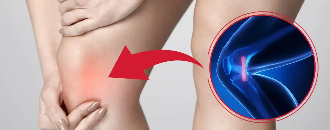
Repetitive Strain Injury (RSI) Explained


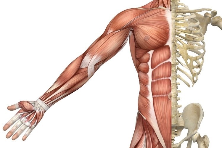
RSI is typically defined as an overuse disorder- a gradual build-up of overload to nerves, tendons, and muscles arising from repetitive movements or activities. Repetitive use of the same motions leads to inflammation and damage to these soft tissues. This disorder mostly affects the upper limb- particularly the elbows, hands and wrists.


Causes

Possible causes of RSI include but are not limited to:

  • Undertaking the same and repetitive movements and stressing the same muscle groups
  • Working in cold environments
  • Assuming a sustained and/or awkward posture for prolonged periods of time
  • Undertaking a particular activity for prolonged periods of time with no rest-breaks
  • Frequent and prolonged use of vibrating equipment
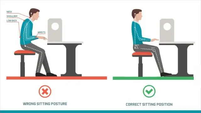
  • Adopting poor postures from working at inappropriately designed workstations
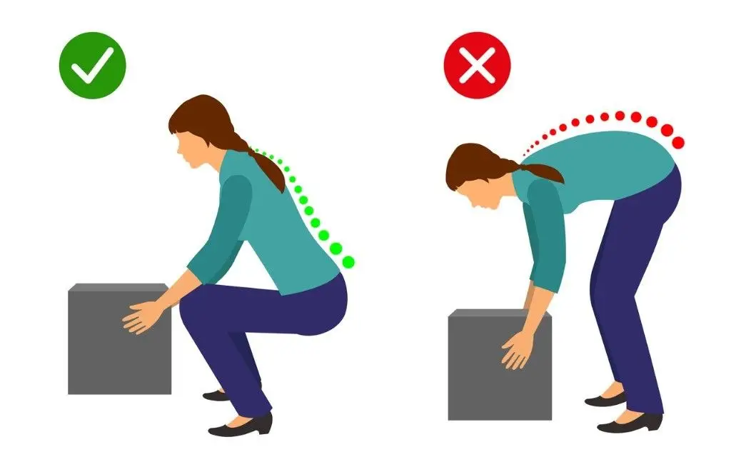
  • Undertaking a motion which involves carrying and/or lifting heavy items


Symptoms

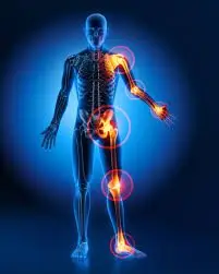
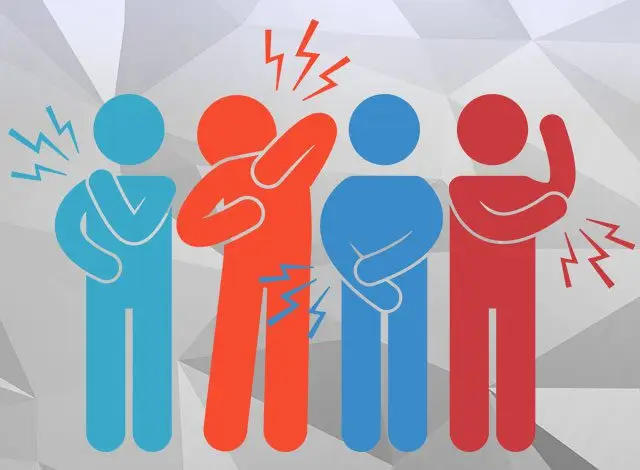
RSI leads to a gradual development of a broad variety of symptoms, which range from mild to severe in severity. RSI particularly affects the muscles and joints of your wrists, hands, elbows, forearms, shoulders, neck. Having said this, RSI can affect other areas of the body as well.

Common symptoms may include:

  • Pain
  • Tingling
  • Cramping
  • Increased sensitivity to heat and cold
  • Tenderness
  • Fatigue
  • Loss of strength
  • Throbbing
  • Soreness
  • Achiness
  • Stiffness
  • Struggling with typical activities of daily living, such as gripping and twisting motions, carrying light weights, writing, kitchen prepping, dressing, personal cares etc

You may develop these symptoms when you undertake a task repetitively for a period of time, and can settle when you stop. Symptoms may settle over a few hours or over the course of a few days. However, if left untreated or is poorly managed, a minor RSI may gradually progress to a nasty chronic injury.


Diagnosis


If you experience mild discomfort whilst completing particular activities at home or at your job, it is a good idea to see your GP or physiotherapist to talk about RSI. But an RSI is not always simple to diagnose as there is no particular clinical test for it. Your GP will enquire about your medical history, occupation and work environment, and other activities to attempt to identify any repetitive motions you undertake that may be the cause of your symptoms. A physical examination will be undertaken, where they will assess your movement, check for pain, inflammation, sensation, tenderness, strength and reflexes in the impacted body part. RSI may be triggered by specific health disorders like bursitis, carpal tunnel, tigger finger, ganglion cyst, or tendonitis (inflammation in your tendons). Your GP can refer you on further diagnostic tests such as X-rays, Ultrasounds, blood tests, MRIs, nerve conduction tests etc, to determine if these underlying disorders may be the cause of your symptoms. You may be also be referred onto a physiotherapist and acupuncturist for conservative treatment and management for mild-moderate issues. If symptoms persist, you will then be referred onto a specialist.


Management

Initial treatment options for the management of RSI symptoms is conservative. This includes:

  • Rest, Ice, Compression, and Elevation (RICE principles)
  • Taking regular breaks between tasks and looking after your posture
  • Undertaking your activities and movements with appropriate form and posture
  • Intake of Nonsteroidal anti-inflammatory drugs (NSAIDs), both oral and topical as prescribed by the GP
  • Use of cold and heat to the impacted area
  • Administration of steroid injections into inflamed joints and tendons
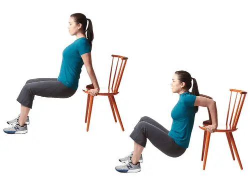
  • Tailored exercise prescription from physiotherapists to correct posture and strengthen and stretch affected muscles
  • Acupuncture
  • Stress reduction and relaxation training
  • Use of splints and braces to help protect and rest the affected muscles and tendons

Ergonomically appropriate adjustments to your workstation and work environment may be recommended by your physio and GP- for example resetting your desk and chair if you’re working at computer, and alterations to your equipment and activities/motions to lessen the strain and stress on your muscles and joints. Surgery may be necessary in some cases.


Prevention

Minimizing repetitive actions particularly if they involve the use of heavy machinery or vibration. Improving your working posture and work-environment as well a taking regular breaks. Employers often undertake risk-assessments when you join a company to determine that the work area is ergonomically fit, comfortable and appropriate for you. You may be able to request for an assessment if you have not had one or are having issues with your work environment

Osteoporosis


Osteoporosis is a condition which results in weak and brittle bones- to such degree that a fall or even mild stresses like coughing or bending over may result in a fracture. Bones are living tissues which are continually being broken down and replaced. However, your bones become osteoporotic when the formation of new bone does not keep up with the loss of old bone. This condition typically develops over time without any pain or other major symptoms, and is generally not diagnosed until you have sustained a fracture. The hip, pelvis, upper arm, spine and wrists are the most common structures affected by osteoporosis- related fractures.


How do you know if you have Osteoporosis?


Because there are no obvious early warning signs and symptoms, it is difficult to pre-diagnose osteoporosis. You may be unaware that you have this condition perhaps till you have one of the following:

  • Sustained a fracture from an incident more easily than you should have- like a simple fall or a bump
  • A decrease in the height of your spinal vertebrae over time
  • Change in posture – stooping or bending forwards
  • Back pain, due to a fractured or collapsed vertebra

Please see your doctor if you experience the following:

  • If you are over the age of 50 and have sustained a fracture
  • Sustained a spine, wrist, or hip for the first time
  • Sustained a fracture more easily than you should have (a simple fall or after a slight bump)


Risk factors

Key factors which may increase your risk of developing osteoporosis include:

  • Females- particularly post-menopausal Caucasian and Asian women
  • Over the age of 50
  • Excessive consumption of caffeine or alcohol
  • Smoking
  • Having a smaller or petite body frame
  • Poor physical activity levels and leading a very sedentary lifestyle
  • Family history of osteoporosis
  • Having low levels of vitamin D and poor dietary calcium intake
  • Decreasing levels of testosterone with ageing in men
  • Estrogen deficiency in women (irregular periods, early (before turning 40) or post-menopausal, surgical removal of the ovaries)
  • Use of long-term medication such as thyroid and epilepsy medications, corticosteroids
  • Having medical conditions such as gastrointestinal diseases; endocrine diseases; rheumatoid arthritis; cancer; and blood disorders


How will you be diagnosed?

Your doctor will review your signs and symptoms, family and medical history. You may be referred on for a specialized X-ray or CT scan to evaluate the bone density to help diagnose osteoporosis. Your bone density will be classified by comparing it to the typical bone density for a person of equivalent gender, size, and age.


How is Osteoporosis treated?

The treatment pathway chosen for the management of this condition is dependent on results of your bone density scan, gender, age, medical history and severity of the condition. Potential treatments for osteoporosis may include exercise, making positive lifestyle changes, vitamin and mineral supplements, and medications. Please consult your doctor for appropriate advice and treatment options.


How can Physiotherapy help?


Your physiotherapist will help you strengthen your bones and your muscles through a personalized and graduated rehabilitation program. Components of this rehabilitation program may include weightbearing aerobic exercises, resistance training using free weights/resistance bands/bodyweight resistance, and exercises to enhance posture, balance and body strength. Your physiotherapist will work with you to find activities that suit your needs and as per your physical activity level.


Managing your Medial Knee Pain: MCL injuries

What is it?

Although your knee has free movement going forwards and backwards, its’ sideward movements are restricted by the robust collateral ligaments on either sides of your knee. The medial collateral ligament (MCL) is situated on the inner part of your knee, but on the outside of your joint. The MCL connects the top of your shinbone (tibia) to the bottom of your femur (thighbone). It helps hold your bones together, provides stability and prevents your knee from bending sideways away from your body.

Injuries to the MCL are from the result of a direct blow to the outer part of your knee- and is most commonly seen in contact sports such as football and soccer. These injures may either over-stretch or cause a tear in the ligament. Whilst surgery may be needed in some severe cases, it is not always the go-to form of management.

Read on to know how physiotherapy can help manage your MCL related-knee pain.


Mechanism of Injury

Injury to the MCL typically occurs when a force drives the lower leg in a sideward direction away from your upper leg and body. Awkwardly landing from a height, twisting of your knee with your foot fixated to the ground, or from a direct blow to the outer part of your knee- most commonly seen in contact sports, are frequent causes of injury to the MCL.


Grading of MCL Injuries

MCL injuries are often graded using the system below:

Grade 1: Regarded as a minor injury- means that the MCL has been overstretched but not torn

Grade 2: Regarded as a moderate injury- means that there is a partial tear in MCL, and presents with some degree of instability in the knee

Grade 3: Regarded as a severe injury- means that the MCL has completely ruptured/torn, and presents with noticeable joint instability


Often 3 MCL injuries are associated with concurrent medial meniscus and ACL ligament damage, which may need surgical intervention. But, the good news is that most MCL injuries may be treated well with conservative physiotherapy management. It usually takes between 2-8 weeks for Grade 1 and 2 MCL injuries to heal, and a graduated rehabilitation programme is highly commended for prevention of future injury.


Signs and Symptoms

Because injury to the MCL may present with similar symptoms as with other knee injuries such as ACL damage, it is vital to have a medical professional such as your physiotherapist evaluate your injury.

Common symptoms of an MCL injury may include:

  • Tenderness and pain along in the inner part of your knee
  • Swelling in the knee
  • Experience catching and locking sensations in the knee joint
  • A ‘pop’ sound at the time of injury
  • Actual or feeling of giving way of the knee (often indicate grade 2 or 3 injury)


Diagnosis

Your physiotherapist will discuss your injury and its presenting symptoms, past medical history (including a history of any prior knee injuries) and will also undertake a thorough physical examination. During the physical examination, your physiotherapist will assess the structures of your injured knee and compare them to the non-injured side. The range of motion, strength and stability of your knee will be assessed. You may be referred on for imaging such as X-rays and Ultrasounds to help aid the diagnosis. For more severe MCL injuries, and if your symptoms do not resolve with conservative physiotherapy management, you may be referred onto a specialist who may consider referring you for an MRI to get a deeper look at your knee.


Management

The management options for MCL injuries will be dependent on the severity of the injury. In the initial stages of injury, management is focused on controlling swelling and pain, whilst allowing your body to initiate healing processes via inflammation. This is typically achieved through the P.O.L.I.C.E. principles (Protect, Optimal Loading, Ice, Compression and Elevation).

Over the counter medication such as ibuprofen and paracetamol may be taken to reduce pain. Other stronger painkillers and NSAIDs may be prescribed by your doctor to help reduce swelling and inflammation as well.

After assessing your knee, your physiotherapist will frame a rehabilitation programme with exercises tailored to your needs. The purpose of physiotherapy is to help restore your knee’s range of motion, stability and strength, which in turn will then allow you to safely return to your usual day-to-day and sporting activities as soon as possible.

Management of most MCL injuries usually only involves knee bracing and physiotherapy treatment. However, in some cases, surgery may be recommended. Particularly if there is damage to more than one ligament or structure in your knee or if you continue to experience instability in spite of physiotherapy.

Dealing with Rheumatoid Arthritis

Rheumatoid arthritis (RA), a chronic inflammatory condition associated with swelling, pain, fatigue, and joint deformity. Although there are no known cures for this condition at present, a combination of treatments are available to help manage your symptoms. RA is the 2nd most common form of arthritis after osteoarthritis and is known to affect 1–2% of New Zealand’s population.


Signs and Symptoms

RA may develop very quickly or gradually over time, with its signs and symptoms, as well as the severity varying from one person to another. This condition is associated with episodes of remission and flare ups, with or without apparent triggers.

Other symptoms may include


  • Swollen, tender joints- (often accompanied by warmth and redness)
  • Joint pain
  • Joint stiffness which worsens in the mornings and after a period of inactivity
  • Fever, loss of appetite weakness, and fatigue
  • Muscle pain
  • Changes to the skin and nails

In the early stages of RA, you may notice its impact on your smaller joints- especially in your toes and fingers. And as this condition develops, your symptoms typically branch out to the bigger joints- your shoulders, ankles, knees, wrists, hips and elbows. Symptoms are likely to affect your joints bilaterally. Over time, RA also causes joints to deform and shift out of place.

Because RA is a systemic condition, it is estimated that approximately 40% of the RA population may experience symptoms and signs other body systems than the joints. These may include:

  • Kidneys, lungs, heart
  • Skin, eyes, mouth
  • Bone marrow
  • Nerves and blood vessels


Causes and Risk Factors

Your immune system is designed to help protect your body from infection and disease. However, in RA, changes occur in your immune system that (for poorly understood reasons), causes it to mistakenly attack the healthy soft-tissues of joints resulting in pain, swelling and inflammation. Because of this ongoing process, over time damages to the lining of your joints and other soft-tissues may lead to bone erosion and joint deformity. It can also have an impact on your heart, lungs, nerves, eyes and skin.

One can get RA at any age, although it is more probable to develop in those in the age bracket of 25-50 years old. Though rare, under 16s may also develop Juvenile RA or Still’s disease.

Risk factors for the development of RA include:

  • Family history of RA
  • Age bracket of 25-50 years old
  • Smoking
  • Women are more likely to develop RA than men
  • Obesity


Diagnosis


At present there is no single test to confirm a clinical RA diagnosis. It is often difficult to differentiate this condition in its initial stages from other forms of connective tissue inflammation (fibromyalgia, lupus, gout etc.).

Your doctor will get your full medical history (as well as any familial history of RA), discuss your signs and symptoms, undertake a physical assessment- particularly of your joints, and refer you on for imaging and blood tests. X-rays may help evaluate RA progression in your joints over time, whilst MRI and ultrasound imaging may help evaluate the severity of RA in your body. The blood test will evaluate your level of anti-bodies and proteins (including the rheumatoid factor protein that is present in approximately eighty percent of the RA population), and markers of inflammation.


Management

At present, though there is no cure for RA, a range of treatments are available which may help slow its’ progression and reduce pain and inflammation, minimise and/or prevent joint damage and maximise joint movement.

A combination of prescribed medication as advised by your doctor and other treatment options as noted below are recommended:

  • Cease smoking if you are smoker
  • Physiotherapy will help improve and maintain your joint range of motion, increase your muscle strength, and decrease your pain. Additionally, your physiotherapist or occupational therapist will be able to teach you ways of using your body efficiently to reduce stress on your joints
  • Finding a balance between rest and activity
  • Use of heat and cold packs to help ease pain and inflammation
  • The use of splints or braces for joint support as needed
  • Hydrotherapy- exercising in water reduces the pressure on your joints, whilst the warmth of the water will relax your muscles and help lessen your pain.
  • Seeking regular medical advice and check-ups to monitor your RA symptoms and the progression of the condition
  • Adopting a healthy and active lifestyle

Managing Your Osteoarthritis

Osteoarthritis impacts millions of people worldwide and is typically known as the most common form of arthritis. It is associated with the wear and tear of the protective cartilage which cushions the ends of your bones in your joints over time. Though this condition may cause damage to any joint in the body, osteoarthritis primarily impacts the joints in your spine, hands, hips, and knees.


Causes and Risk factors

Over time, the gradual deterioration of the cartilage which cushions the ends of your bones in your joints causes arthritis. Cartilage is a solid slippery tissue which allows almost frictionless joint movement. As the cartilage wears down, bone will eventually rub on bone.

This condition is typically characterized as a wear and tear degenerative disorder. However, in addition to the breakdown of the cartilage, it also impacts the joint as a whole. Osteoarthritis triggers alterations in the bone and damages connective tissue which holds the joint together and attaches your muscles to your bones. Inflammation of the lining of the joint is also triggered.

Factors which may put you at higher risk of developing osteoarthritis include but are not limited to:

  • Your age- the risk increases with getting older
  • Gender- though unclear why, but women are more perceptible to developing osteoarthritis
  • Bony deformities- those with abnormal joints or defective cartilage
  • Sustaining bony or joint injuries like those which take place during sport or from an accident.
  • The risk increases with obesity- the more you weigh, the greater your risk, as it adds more stress to your weight-bearing joints (particularly hips and knees)
  • Your occupation or a sport that you play which puts repetitive and excessive stress/loading on the joints, can eventually lead to the development of osteoarthritis.
  • Certain co-morbidities such as diabetes


Common symptoms

Below are some common examples of symptoms you may experience with arthritis. These may develop and worsen gradually over time

  • Pain: Your joints may hurt before and/or after undertaking an activity
  • Loss of joint range of motion– loss of overall joint flexibility and movement
  • Tenderness felt on applying light pressure to the joint
  • Joint stiffness that is most noticeable on waking up first thing in the morning or after a prolonged period of inactivity
  • Noticeable changes in joint pain with changes in the weather- particularly colder weathers
  • Sensations of grating and grinding// sounds of clicking and popping (crepitus) when you use the joint
  • You may notice swelling and redness around the joint, which may be triggered by soft tissue inflammation
  • Bony spurs that feel like hard bumps may develop around the impacted joint


How will I be diagnosed?

Osteoarthritis is typically diagnosed based on your medical and occupation history and a physical examination undertaken by your doctor. During the physical examination, your doctor will assess your affected joint(s) for swelling, tenderness, redness, and stiffness. X-rays may be recommended to reveal cartilage loss (the narrowing of the space between the bones of your joints), changes in bone, and bony spurs around the joint. Blood tests may be used to rule out other causes of joint pains like rheumatoid arthritis. Joint fluid analyses may also be used to test for inflammation to ascertain if the pain is triggered by an infection or gout instead of osteoarthritis.


Management

Though there isn’t a cure for osteoarthritis, various treatments which can help relieve symptoms of pain and disability are available.

Lifestyle modifications: Changes to your daily life may protect your joints and slow the progression of osteoarthritis.  Minimising activities which exacerbate your symptoms such as climbing stairs, squatting. Swapping high-impact activities like running and jogging to lower-impact activities such as cycling or hydrotherapy will decrease the stress on your joints. Weight-loss reduces the stress and loading on your joints, which then results in less pain with increased function.

Assistive aids: Using assistive aids like a stick/cane, wearing proper shoes w orthotics and supportive braces/sleeves may improve your stability and support your functional capabilities.

Physiotherapy: Targeted exercises may help improve your flexibility as well as build strength in your muscles. Your physiotherapist will develop a personalised active rehabilitation program which is safe and will meet your requirements and lifestyles.

Medications: Various kinds of medication (such as paracetamol and NSAIDs) maybe helpful in treating and controlling the symptoms of osteoarthritis. As everyone responds differently to medications, your doctor will prescribe medicines (type and dosage), which is safe and will work best for you.

Cortisones: Strong anti-inflammatory agents which is injected into the affected joint to give pain relieve and decrease inflammation for a short period of time. Due to potential side-effects, it may be recommended to restrict the number of injections to 2-3 per year.

Other: Heat and ice applications, self-massaging with pain-relieving creams/ointments and/or wearing elastic supports may provide some relief from your pain and give you support.

Surgery: Surgery may be recommended if there is considerable degeneration in your joints and/or if your osteoarthritic pain causes disability that is not relieved with conservative management. Your doctor or specialist will discuss your options with you.

Meet Your Body Parts!

Here are definitions of common terms for body parts you may hear your doctor or physio use!


Ligaments

Ligaments are cordlike extensions that serve to connect ends of two bones to form a joint. They are made up of strong, durable, slightly elastic bandlike structures comprised of collagen fibres. The structural make up of ligaments is advantageous providing joint stability by limiting excessive movement.

What are Ligaments? (with pictures)

Tendons

Similar to ligaments, tendons contain densely packed bundles of tough collagen fibres that hold muscles together to the bone. They are located at the ends of every muscle in the human body. Bound together in tight sheaths they are made to withstand tension and transmit forces exerted by the muscle to the bone to cause movement.


10 Facts About Tendons | Physio Direct - Rural Physio at Your Doorstep

Muscles

Human body is made up of over 600 muscles categorised into three different types – cardiac, smooth and skeletal muscle.

  • Cardiac muscle – is only found in the walls of the heart. Its contractions help propel blood through the blood vessels to all part of the body.
  • Smooth muscle – is found mainly in the lining of internal organs (except the heart) including digestive and uninary tract organs, blood vessels. Smooth muscle works to transport substances through the organs by alternately contracting and relaxing.
  • Skeletal muscles – Skeletal muscles are the most abundant type of muscles that form the flesh of the body. They are attached to bones of the skeleton by tendons. They are responsible for voluntary movements of body. Facial expression, mobility, postural control and breathing are some of the movements we observe when skeletal muscles are subjected to voluntary control.


Muscle types — Science Learning Hub

Bones

Skeletal system of the human body is made up of 206 bones. Bones are most involved in providing an architectural framework by providing body shape, support and protection of vital organs and for locomotion. Besides these functions, bone is a reservoir for mineral and fats as a source of stored energy and formation of blood cells. Bones are classified by their shape as long, short, flat and irregular. They are connected by ligaments to form joints.

Let's learn about bones | Science News for Students

Cartilage

There are three different types of cartilage found in the human body – hyaline, elastic and fibrocartilage. Hyaline cartilage is the most common cartilage in the human body. It covers the ends of most bones at movable joints, connects ribs to the breastbone, forms the voice-box and nasal passages. It  consists of high water content that provides resilience to withstand great compressive forces found predominantly in joints.


What Is Cartilage?

Ankle injuries – Your first step to recovery

Common? Oh Yes!

The ankle is the most commonly injured joint in sport. This does not exclude other people such as active hikers, beach goers and even your average Sunday stroller. Good news though – your ankle injury is highly likely to be uncomplicated.

It is still vital that your ankle is examined, evaluated and treated early. This will ensure a swift return to activity and prevent further complications.


The road to recovery

Planning for Resiliency and the Road to Recovery

Your clinician will ask you some questions related to how you injured your ankle, pain, instability and any past episodes of injury. The earlier you get your ankle checked, the sooner your recovery will begin.

Keeping a mental note of things like initial pain, swelling, ability to walk and balance will go a long way in assisting your clinician to making an accurate diagnosis.


Investigations

Ankle x-rays

In most cases, initial X – rays are done to rule out broken bones.

Ultrasounds can be used to diagnose some ligament and tendon damage.

MRI is the best form of imaging but this does come at a higher cost and higher exposure to radiation. These are usually done after failed conservative treatment or in instances where pain remains high for longer periods.

A CT scan is helpful with complicated foot and ankle fractures. It will normally be ordered by a specialist surgeon who is planning for an operation.


What to look out for

Ankle sprains:

This is normally a twisting injury that causes a stretch or tear of ligaments surrounding the ankle. Your health care professional will provide you with all the information and tools you need for recovery.

These heal relatively quickly when the outside border of the foot is affected and a little slower when the inside border of the ankle is affected.

You will normally feel pain on certain ankle movements, stiffness in the ankle and experience some swelling and bruising.

Ankle - Wikipedia


Ankle Fractures:

These normally present with swelling, bruising and pain initially – although not always. In some cases, it is too painful to put weight on the ankle.

They are usually best confirmed with X – ray and specialist referral.

Management may be surgical or non-surgical depending on the severity and site of the fracture.

Fractures generally take longer to recover compared to sprains.


What treatment to expect

Foot and Ankle Doctor | EmergeOrtho—Triangle Region

Acute phase:

Your healthcare professional will normally initiate techniques to minimise your pain and swelling with rest, ice, compression and elevation.

Analgesia and anti – inflammatory medication may also be used.

Strapping may be used for stability at this stage and can be done by your physiotherapist.

You will also be encouraged to increase movement and begin strengthening.


Rehabilitation phase:

Balance and proprioceptive exercises will be given to you by your physiotherapist.

Strengthening will continue and running will start soon.

Once running in a linear motion pain free, you will progress to sport specific exercises.

Finally, you will return to sport or previous function such as trekking with a graded program.

Strapping may continue for up to 12 months after your injury in order to prevent re–injury.


What can you do on the day of the injury?

Rest by reducing time spent walking or standing. This will help the ankle to heal.

Ice the ankle for up to 20 minutes every couple of hours.

Compress the ankle with a firm bandage during the day and remove the bandage at night.

Elevate the leg.

Attempt circulatory exercises such as ankle circles and foot pumps (About 10 – 30 repetitions every couple of hours).

Contact your health professional or physiotherapist in order to make appointment for assessment.

If you are unable to stand on your leg or have excruciating pain in the ankle, head on to the local emergency department for immediate investigation.


Remember, your injury will heal and you will recover!


To find your nearest Physio Fusion clinic and book an appointment call 09 6266186 or visit our website https://physiofusion.co.nz

We Want You To Understand Your Pain

What is pain?

Pain is the brain giving out a message to protect you . It is part of our bodies natural defense system.

When an event occurs that we need protecting from, our brains response is to increase our pain. Living things detect and respond to stimulus. | Characteristics of living  things, Natural hairstyles for kids, Characteristics

Example: Putting a hand on a hot pan. You feel pain, which is the brain giving you a warning signal to move your hand away.

Pain is all about protection, never about measuring the condition of the tissues in the body.

In this instance we experience a high level of pain to prevent a bigger injury eg. causing a burn.

This is our bodies way of protecting us

So what is happening in our brain?

  • We as human beings are amazingly adaptable. The longer we have pain, the longer our brains learn to produce pain.

  • It hurts in the tissues (back, knee, hand), but the problem is in the nervous system. There is an adaptation within our nervous system.

Long term pain or Chronic pain

Chronic pain is defined as persistent or recurrent pain lasting longer than 3 months

If you have had pain for more than 3 months, your system is now overprotective. Your nervous system and immune systems have learnt.


Movement is medicine: why exercise therapy reduces chronic pain - Hinge  Health


  • With chronic pain the buffer size is increased- therefore pain comes on quicker than before.

  • You get pain when you are not anywhere near being in danger.

Management of chronic pain:

  • Identify why your brain is protecting you – fear, anxiety, quality of movement, posture, injury

  • Reduce the size of your buffer – desensitization, reassurance, progressive load management

  • Understand your pain- know that it may not go completely, but can become more manageable

Medication:

  • Depending on the type of pain you are experiencing, painkillers/NSAID’s may be beneficial in the short term – speak to your GP/pharmacist

  • Note: the research does not show good outcomes for the long-term use of medication to treat chronic pain.

Occupational Therapy:

  • If you have been off work because of your pain – try to return to work asap. An OT can discuss and plan a gradual return to work plan for you

  • They can also assist by providing aids to helps with daily tasks such as getting out of bed, or putting on shoes.

Physiotherapy:

  • There is no quick fix for persistent pain. We can help guide you along your journey to recovery

  • With persistence and hard work you can learn to adapt and cope with your pain

  • Movement is king – it is critical for retraining the system

  • Movement gradually suppresses the pain system.

  • Finding any form of exercise or movement that you enjoy and gradually increasing the volume over time

  • Moving regularly- on your good days and your bad days

Self Help:

  • Meditation or mindfulness – Apps such as Headspace or Calm are easy to use and will guide you through the process

  • The Pain ToolKit

The myth about foot pronation (flat foot)

Myth: Foot pronation(flat foot) is the enemy.

Quest - Article - Surgery Sometimes, Bracing Often, Caution Always |  Muscular Dystrophy Association

In the foot, pronation should occur naturally when the foot comes into contact with the ground. Pronation will appear as the foot rolling inward and the arch flattening.

What are the benefits?

  • Dissipates the force that the foot receives from the ground

  • Allows the foot to become a stable and mobile adaptor to enhance movement opportunity

  • Loads the muscles of the extensor chain (calf, quads, glutes) to convert ground reaction forces into forward momentum so we can propel efficiently.

So why have I been told this is bad?

So as you are now aware, pronation is very normal and a critical movement to ensure we move and propel ourselves forward efficiently.

What you may have heard someone say to you is that you ‘overpronate’?

Firstly, overpronation is subjective and not as black and white as it is sometimes made out to be. Overpronation has be defined as: ‘a foot that rolls inward toward the arch excessively’.

Foot Pronation: Underpronation & overpronation explained- The Foot Clinic

What we must understand is that a pronation can only happen when the foot has a stable tripod on the ground. This means that the calcaneus (heel bone), 1st metatarsal (big toe knuckle), 5th metatarsal (little toe knuckle) must all remain in contact when the foot rolls inwards and the arch flattens.

The Foot Tripod - Fix Flat Feet

So, If you have been told you are ‘overpronated’ , it is most likely that your whole foot is ‘everting’ NOT ‘overpronating’.

What is Eversion?

Eversion can be defined as: ‘the process of turning inside-out’.

In pronation your heel must naturally ‘evert’ (sole of the heel will move away from the midline of the body) NOT your whole foot.

If your ‘whole foot’ everts (turns out) you will no longer have a stable foot tripod as the 5th metatarsal (little toe) will lose contact with the ground.

The key to ensuring this does not happen is to provide an environment for the bones of the midfoot (middle of the foot) and forefoot (toes) to experience the opposite motion to that of the heel. This will mean that the foot has an opportunity to truly pronate with a tripod on the ground.


So how can you help me do that?

  • At Physio fusion we can help you to bring your own body into alignment and create an environment in which the healing can begin

  • Foot strengthening exercises

  • Footwear advice

  • Referral to other healthcare specialists for further assistance (e.g. podiatrists)

To find your nearest Physio Fusion clinic and book an appointment call 09 6266186 or visit our website https://physiofusion.co.nz

Exercises you can do during lockdown

I know how hard it can be when it comes to working out and its really easy to find an excuse to avoid lockdown workouts but it’s not just about staying in shape its also about keeping active to feel mentally strong. We know that exercise does wonders for mental health and at a time where its all uncertainty, anxiety, and a daily dose of doom and gloom, we all need to work out that frustration somewhere!

If you’re struggling with a lack of motivation or negative mindsets there’s only one way to turn it all around: Take :boom: ACTION :boom:


How much exercise is enough?

You’ve heard the saying something is always better than nothing and that’s the case when it comes to exercise. The current recommendation for adults according to Best Exercises for Health and Weight Loss.org is to aim for 150 minutes of moderate intensity activity every week (or 75 minutes of vigorous intensity) with two sessions of strength building activities per week. That’s about 30 minutes of movement, five times per week.

This is a quick full body routine:

Bodyweight Squats

Stand with your feet shoulder-width apart. Engage your core muscles and gently squat down. As you squat, bend from your hips. Keep your back straight as you push your hips back and counterbalance by leaning your torso forwards. Keep your knees aligned with your toes. Your weight should be evenly on your heels and the balls of your feet, not your toes. It might help to image your are trying to sit down in a chair that is too far away from you. Tense your bottom muscles at the bottom of the squat and keep them tense as you straighten back up to the start position.

Arm dips

Start in a seated position. Place your hands on the seat of the chair and use your arms to move yourself forwards towards the front of the chair. You will need to move your feet further forwards to help your stability. From this position, use the strength of your arms to slowly lower your body directly down towards the floor and then raise yourself back up. Do not actually sit on the floor and keep your hands close in beside you. Relax and repeat.


Lunges

Stand straight with your arms to the side or on your hips. Take a large step forwards on your affected leg, then drop your hips directly down between both feet, bending your hips and knees to a 90 degrees. Push back up to the starting position, and repeat. Make sure you take a large enough step that your front knee does not travel over your foot, and ensure your knee travels directly forwards. Keep your body up straight throughout the movement.


Mountain Climbers

Adopt a plank position insuring your hands are directly beneath your shoulders.
Fully flex one hip and hold.
Extend the bent leg to the rear and repeat the movement pattern on the opposite side