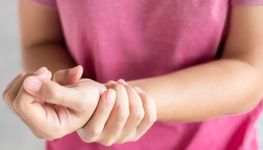
De Quervains tenosynovitis is a painful condition caused by inflammation of two prominent tendons that are located at the wrist and thumb.
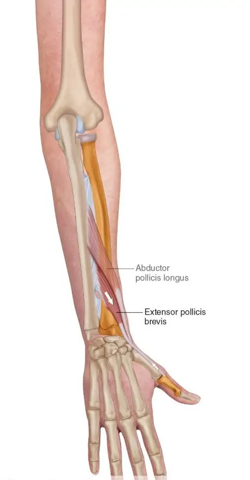
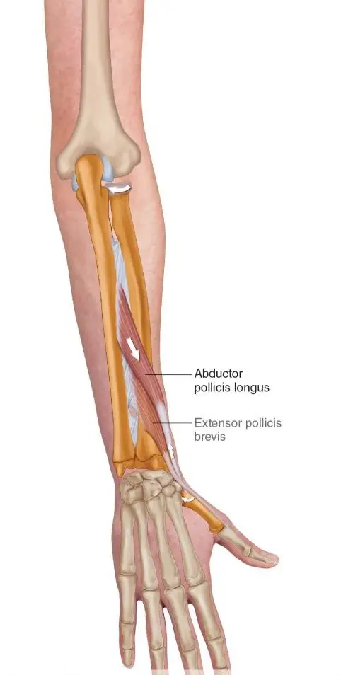
The two tendons called the Extensor pollicus brevis and Abductor pollicus longus originate from the middle of the forearm travel down towards and over the wrist to insert into the thumb. Collectively they function to extend the thumb, whilst abductor pollicus longus extends and also abducts the thumb (lifting thumb up to the ceiling).
What causes it?
The most common cause of De Quervains tenosynovitis is the repetitive overuse of thumb and wrist whether it is occupational or hobby related. For example, the repetitive thumb movement whilst using scissors by hair dressers, landscapers using shears or whilst gardening). Trauma to the tendons from injuries to the wrist or the thumb can cause inflammation of the tendons.
In some cases, age related degeneration of the tendon sheath or underlying conditions such as rheumatoid arthritis increases the risk of the developing De Quervains tenosynovitis. Hormonal changes resulting in fluid build up in young mothers can commonly result in De Quervains tenosynovitis.
Symptoms
Commonly your symptoms may include:
Pain located at base of your thumb
Pain elicited by movement of thumb (gripping or making a fist)
Grating or snapping feeling
Tightness in the wrist
Swelling surrounding the base of thumb and wrist
How is De Quervains tenosynovitis diagnosed?
Your doctor or physiotherapist will be able to diagnose the condition based on your symptoms and after doing a thorough movement assessment to rule out any other potential diagnosis.
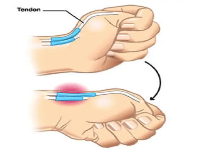
Finkelstein test is used to elicit symptoms to confirm De Quervains tenosynovitis.
How to test:
Wrap your thumb with your fingers.
Slowly bend your wrist down
A positive test would elicit pain at the site of the two tendons.
Radiological investigations in lights of ultrasound and an x-ray might be recommended for further investigations, particularly to confirm clinical diagnosis or to rule out any other possible causes of De Quervains such as osteoarthritis.
What treatment options are available?
Conservative (non-surgical) management
Conservative management measures are generally recommended as the first line of management for mild to moderate symptoms. This is because up to 60-70% of symptoms are likely to improve over a period of 6-8 weeks of regular physiotherapy intervention. In this period, the following strategies are recommended by your therapist to fast-track your recovery
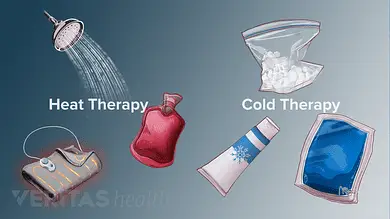
Rest and application of heat or cold packs
Avoid repetitive use of thumb
Pain medications (anti-inflammatory medications) such as diclofenac or ibuprofen
Splints or braces
Steroid injection
Surgical management
In more severe cases when conservative management has failed, surgery may be recommended by an orthopaedic specialist or surgeon.
Prior to your surgery you will have the opportunity to thoroughly discuss with your surgeon the details of the surgical procedure and about the post operative rehabilitation process.
Surgical procedure
Surgery may be performed under general or local anaesthesia. A small incision is made at the wrist and thumb region. The covering of the tendons (sheath) is then separated and expanded to provide the tendon space to allow the tendon to move smoothly within the sheath. After this the, the incision in then sutured with a firm dressing applied over the suture site.
While you recover from the surgery, an information sheet with post operative guidelines will be provided to you by your surgical team. It is important that you must follow the guidelines recommended by your surgeon for optimal recovery.
In most cases your will have a follow up with your surgeon few weeks after your surgery to check your wound healing and your progress. You are often times referred to physiotherapy for strength and conditioning of your wrist and hand movements to facilitate your recovery.
Sciatica is not a diagnosis, it is a term that describes symptoms of pain, pins and needles, numbness and in some cases weakness that radiates along path of the sciatic nerve from the lower back to buttocks and leg.
Causes of Sciatica?
Disc derangement / herniation Disc injuries are most common cause of sciatica. Discs are cushion like pads located between each spinal segments that act as shock absorbers. The core interior of the discs is made up a gel like substance called the nucleus pulposus surrounded by thick fibrous outer ring called the annulus. Sudden forces applied to the disc can result in the the core interior to push through the outer ring resulting in a disc bulge or in severe cases can rupture the outer ring resulting in disc herniation which can compress the nerve root.
Disc degeneration / Arthritis / stenosis
Age related degenerative changes in the spine can result thinning of the disc and narrowing of the spinal joints. Overtime the narrowing results in bony growths that can compress the nerve roots resulting in sciatica.
Soft tissue injury resulting in inflammation
Thick ligaments and connective tissue envelope the spinal segments to optimize stability. Injury to any of the structures will result in inflammation and swelling which can affect the sciatic nerve directly resulting in sciatic symptoms.
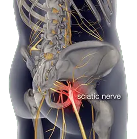
Piriformis syndrome
Piriformis muscle is a located deep in the buttock region. It originates from the sacrum and inserts into the upper part of the hip. The sciatic nerve travels adjacent to the piriformis muscles. Injury to the muscle resulting in tightness or spasm directly affects the sciatic nerve resulting in symptoms.
Other possible causes:
Sacroiliac Joint Dysfunction
Hip joint injury or arthritis
Spinal fractures
Tumors
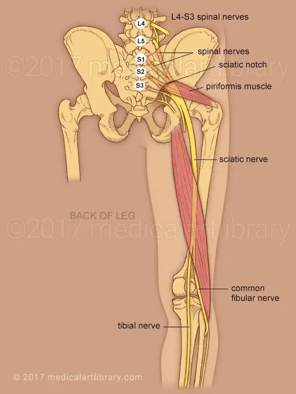
Anatomy of the Sciatic nerve
Sciatic nerve is the largest nerve in the human body. It originates in the lower back from five branches of nerves that extend from the spinal cord. The branches exit the spine at nerve roots L4, L5, S1, S2, S3 connect together to form the sciatic nerve.
The large sciatic nerve then travels deep in gluteal region and descends vertically down to the back of the thigh. It supplies motor function and sensation to the skin and all muscles in the posterior compartment of thigh.
At the knee joint the sciatic nerve then divides into two branches the tibial nerve and common fibular nerve.
What exactly does it feel like?
Symptoms of Sciatica are often characterized by one or more of the following features:
Unilateral. Sciatica is typically affects one leg.
Pain. Nature of pain is often constant with heaviness or dull ache. You may experience sharp, shooting, electric shocks intermittently with postural movements.
Neural irritation. pins and needles with occasional postural numbness is common. Postural numbness can occur when you sit or stand for a period of time, but should resolve with movement. However, if numbness is constant you must be reviewed by your general practitioner or your physiotherapist.
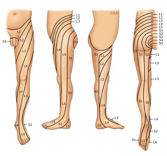
Location of pain: symptoms of sciatica are felt along the path of the large sciatic nerve. The following picture shows the potential pathways you may feel your symptoms radiate to depending on the origin of nerve irritation or entrapment. Most commonly the areas affected included the lower back, lateral thigh extending to the calf and foot.
If symptoms are presented on both sides with additional symptoms outlined below – this could warrant an urgent medical review.
Red flags
Signs and symptoms that require prompt medical assessment include:
Age >50 years
History of trauma
Severe unrelenting pain that does not resolve with rest or pain control
Partial or complete loss of bowel and bladder function or control
Numbness in private regions and the affected side of leg or both
Discoloration of skin in comparison to unaffected side
Recent or current infection with fever chills and night sweats
Sudden unplanned weight-loss
History of cancer, kidney dysfunction
Diagnosis
It is important to correctly identify the cause your sciatica is essential in order to formulate an effective treatment plan to manage your symptoms and improve function.
Your GP or a physiotherapist will conduct a thorough diagnostic assessment. Your consultation with your clinician will begin with a comprehensive conversion that allows your clinician to formulate an understanding around potential causes of your symptoms. This includes questions specific to your presenting concerns, general health, history of injuries contributing and medication history and your symptoms management strategies. A physical examination is then followed where by your clinician will assess the range of movement of your lower back and lower extremities, reflexes, strength and sensation assessment to test the integrity of the nerve.
Radiographic examination
Further diagnostic examination in forms of radiographic imaging may be recommended by your clinician to assess the quality of your joints, alignment and check for the presence of any potential lesions contributing to your symptoms.
XRAY – commonly used in initial stages to review underlying joint pathology such as wear and tear of joints, fractures or in some cases to view lesions or tumors
MRI – high standard imaging that is able to examine in very refined detail possible soft tissues such as muscles, ligaments and internal organs as well as the bony architecture and possible disc injuries.
Discogram – A discogram test may be helpful in determining abnormalities in an intervertebral disc. A contrast dye injected into the tissues may allow abnormalities in the disc, such as bulging or herniation to be seen on a medical imaging scan (such as computed tomography scan).
Treatment
It is advisable to treat sciatica as early as possible in order to avoid the progression of symptoms. Treatments for sciatica may include both non-surgical and surgical approaches.
Typically, non-surgical management is recommended first. Surgery may be required if non-surgical methods have failed to manage your symptoms or the underlying cause is causing deterioration of symptoms. However, in a few severe cases where red flags are presented, surgery may be considered as the first option
Non-surgical approach is the first step to management. This includes intake of pain medications as prescribed by your doctors and referral to physiotherapy.
Pain medications
Your doctor will prescribe pain medications best suited for your symptoms. These may include
Non-steroidal anti-inflammatory medications such as ibuprofen, celecoxib
Neuropathic medications such as gabapentin, amitriptyline
Analgesics such as codeine, tramadol or oxycodone.
Muscle relaxants such as norflex
Physiotherapy
Physiotherapy will incorporate a combination of gentle strengthening, stretching, and the use of manual therapy to facilitate therapeutic gains.
The goals of physiotherapy for sciatic symptom management includes:
Strengthen muscles of the spine, core and lower extremities.
Improve flexibility of any tight muscles
Improve mobilization of the sciatic nerve
Facilitate optimal circulation through slight conditioning exercise (walking, swimming)
Education around activity modifications (especially for work-related participation)
Alternative therapies such as acupuncture may be recommended in combination to physiotherapy to facilitate management of your symptoms.
Acute mild sciatica usually improves with 4 to 6 weeks with regular conservative treatment. However, for moderate to severe cases of sciatica especially with a chronic underlying pathology pain may last over 8 weeks and, treatment time may take longer.
Steroid Injections
Steroid injections are slightly an invasive method used for pain management. Your specialist or an orthopedic surgeon may recommend and administer the injection. In addition to this, injections are also used as a diagnostic method to identify the target nerves or nerve roots affected. The common types of injections for sciatic pain relief include epidural injections.
Surgical approach
In cases where pain and or weakness persists for more than 6-8 weeks or if your symptoms are affecting everyday activities – Surgery may be considered. Your physiotherapist or doctor will arrange the referral for you to meet with an orthopedic back surgeon. Depending on the cause of your sciatica, your surgeon will discuss with you in detail the intended surgical approach, risks involved, post operative management and possible adverse reactions you may have after surgery.
Osteoarthritis impacts millions of people worldwide and is typically known as the most common form of arthritis. It is associated with the wear and tear of the protective cartilage which cushions the ends of your bones in your joints over time. Though this condition may cause damage to any joint in the body, osteoarthritis primarily impacts the joints in your spine, hands, hips, and knees.
Causes and Risk factors
Over time, the gradual deterioration of the cartilage which cushions the ends of your bones in your joints causes arthritis. Cartilage is a solid slippery tissue which allows almost frictionless joint movement. As the cartilage wears down, bone will eventually rub on bone.
This condition is typically characterized as a wear and tear degenerative disorder. However, in addition to the breakdown of the cartilage, it also impacts the joint as a whole. Osteoarthritis triggers alterations in the bone and damages connective tissue which holds the joint together and attaches your muscles to your bones. Inflammation of the lining of the joint is also triggered.
Factors which may put you at higher risk of developing osteoarthritis include but are not limited to:
Your age- the risk increases with getting older
Gender- though unclear why, but women are more perceptible to developing osteoarthritis
Bony deformities- those with abnormal joints or defective cartilage
Sustaining bony or joint injuries like those which take place during sport or from an accident.
The risk increases with obesity- the more you weigh, the greater your risk, as it adds more stress to your weight-bearing joints (particularly hips and knees)
Your occupation or a sport that you play which puts repetitive and excessive stress/loading on the joints, can eventually lead to the development of osteoarthritis.
Certain co-morbidities such as diabetes
Common symptoms
Below are some common examples of symptoms you may experience with arthritis. These may develop and worsen gradually over time
Pain: Your joints may hurt before and/or after undertaking an activity
Loss of joint range of motion– loss of overall joint flexibility and movement
Tenderness felt on applying light pressure to the joint
Joint stiffness that is most noticeable on waking up first thing in the morning or after a prolonged period of inactivity
Noticeable changes in joint pain with changes in the weather- particularly colder weathers
Sensations of grating and grinding// sounds of clicking and popping (crepitus) when you use the joint
You may notice swelling and redness around the joint, which may be triggered by soft tissue inflammation
Bony spurs that feel like hard bumps may develop around the impacted joint
How will I be diagnosed?
Osteoarthritis is typically diagnosed based on your medical and occupation history and a physical examination undertaken by your doctor. During the physical examination, your doctor will assess your affected joint(s) for swelling, tenderness, redness, and stiffness. X-rays may be recommended to reveal cartilage loss (the narrowing of the space between the bones of your joints), changes in bone, and bony spurs around the joint. Blood tests may be used to rule out other causes of joint pains like rheumatoid arthritis. Joint fluid analyses may also be used to test for inflammation to ascertain if the pain is triggered by an infection or gout instead of osteoarthritis.
Management
Though there isn’t a cure for osteoarthritis, various treatments which can help relieve symptoms of pain and disability are available.
Lifestyle modifications: Changes to your daily life may protect your joints and slow the progression of osteoarthritis. Minimising activities which exacerbate your symptoms such as climbing stairs, squatting. Swapping high-impact activities like running and jogging to lower-impact activities such as cycling or hydrotherapy will decrease the stress on your joints. Weight-loss reduces the stress and loading on your joints, which then results in less pain with increased function.
Assistive aids: Using assistive aids like a stick/cane, wearing proper shoes w orthotics and supportive braces/sleeves may improve your stability and support your functional capabilities.
Physiotherapy: Targeted exercises may help improve your flexibility as well as build strength in your muscles. Your physiotherapist will develop a personalised active rehabilitation program which is safe and will meet your requirements and lifestyles.
Medications: Various kinds of medication (such as paracetamol and NSAIDs) maybe helpful in treating and controlling the symptoms of osteoarthritis. As everyone responds differently to medications, your doctor will prescribe medicines (type and dosage), which is safe and will work best for you.
Cortisones: Strong anti-inflammatory agents which is injected into the affected joint to give pain relieve and decrease inflammation for a short period of time. Due to potential side-effects, it may be recommended to restrict the number of injections to 2-3 per year.
Other: Heat and ice applications, self-massaging with pain-relieving creams/ointments and/or wearing elastic supports may provide some relief from your pain and give you support.
Surgery: Surgery may be recommended if there is considerable degeneration in your joints and/or if your osteoarthritic pain causes disability that is not relieved with conservative management. Your doctor or specialist will discuss your options with you.