fbpx
Your first step to recovery

Osteoporosis


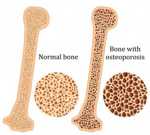
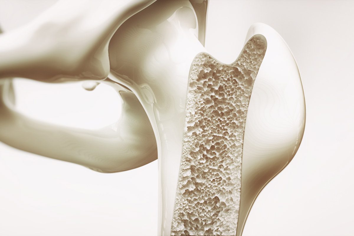
Osteoporosis is a condition which results in weak and brittle bones- to such degree that a fall or even mild stresses like coughing or bending over may result in a fracture. Bones are living tissues which are continually being broken down and replaced. However, your bones become osteoporotic when the formation of new bone does not keep up with the loss of old bone. This condition typically develops over time without any pain or other major symptoms, and is generally not diagnosed until you have sustained a fracture. The hip, pelvis, upper arm, spine and wrists are the most common structures affected by osteoporosis- related fractures.


How do you know if you have Osteoporosis?


Because there are no obvious early warning signs and symptoms, it is difficult to pre-diagnose osteoporosis. You may be unaware that you have this condition perhaps till you have one of the following:

  • Sustained a fracture from an incident more easily than you should have- like a simple fall or a bump
  • A decrease in the height of your spinal vertebrae over time
  • Change in posture – stooping or bending forwards
  • Back pain, due to a fractured or collapsed vertebra

Please see your doctor if you experience the following:

  • If you are over the age of 50 and have sustained a fracture
  • Sustained a spine, wrist, or hip for the first time
  • Sustained a fracture more easily than you should have (a simple fall or after a slight bump)


Risk factors

Key factors which may increase your risk of developing osteoporosis include:

  • Females- particularly post-menopausal Caucasian and Asian women
  • Over the age of 50
  • Excessive consumption of caffeine or alcohol
  • Smoking
  • Having a smaller or petite body frame
  • Poor physical activity levels and leading a very sedentary lifestyle
  • Family history of osteoporosis
  • Having low levels of vitamin D and poor dietary calcium intake
  • Decreasing levels of testosterone with ageing in men
  • Estrogen deficiency in women (irregular periods, early (before turning 40) or post-menopausal, surgical removal of the ovaries)
  • Use of long-term medication such as thyroid and epilepsy medications, corticosteroids
  • Having medical conditions such as gastrointestinal diseases; endocrine diseases; rheumatoid arthritis; cancer; and blood disorders


How will you be diagnosed?

Your doctor will review your signs and symptoms, family and medical history. You may be referred on for a specialized X-ray or CT scan to evaluate the bone density to help diagnose osteoporosis. Your bone density will be classified by comparing it to the typical bone density for a person of equivalent gender, size, and age.


How is Osteoporosis treated?

The treatment pathway chosen for the management of this condition is dependent on results of your bone density scan, gender, age, medical history and severity of the condition. Potential treatments for osteoporosis may include exercise, making positive lifestyle changes, vitamin and mineral supplements, and medications. Please consult your doctor for appropriate advice and treatment options.


How can Physiotherapy help?


Your physiotherapist will help you strengthen your bones and your muscles through a personalized and graduated rehabilitation program. Components of this rehabilitation program may include weightbearing aerobic exercises, resistance training using free weights/resistance bands/bodyweight resistance, and exercises to enhance posture, balance and body strength. Your physiotherapist will work with you to find activities that suit your needs and as per your physical activity level.



Dealing with Rheumatoid Arthritis

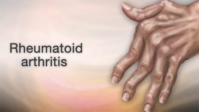
Rheumatoid arthritis (RA), a chronic inflammatory condition associated with swelling, pain, fatigue, and joint deformity. Although there are no known cures for this condition at present, a combination of treatments are available to help manage your symptoms. RA is the 2nd most common form of arthritis after osteoarthritis and is known to affect 1–2% of New Zealand’s population.


Signs and Symptoms

RA may develop very quickly or gradually over time, with its signs and symptoms, as well as the severity varying from one person to another. This condition is associated with episodes of remission and flare ups, with or without apparent triggers.

Other symptoms may include


  • Swollen, tender joints- (often accompanied by warmth and redness)
  • Joint pain
  • Joint stiffness which worsens in the mornings and after a period of inactivity
  • Fever, loss of appetite weakness, and fatigue
  • Muscle pain
  • Changes to the skin and nails

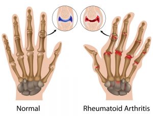
In the early stages of RA, you may notice its impact on your smaller joints- especially in your toes and fingers. And as this condition develops, your symptoms typically branch out to the bigger joints- your shoulders, ankles, knees, wrists, hips and elbows. Symptoms are likely to affect your joints bilaterally. Over time, RA also causes joints to deform and shift out of place.

Because RA is a systemic condition, it is estimated that approximately 40% of the RA population may experience symptoms and signs other body systems than the joints. These may include:

  • Kidneys, lungs, heart
  • Skin, eyes, mouth
  • Bone marrow
  • Nerves and blood vessels


Causes and Risk Factors

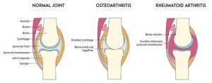
Your immune system is designed to help protect your body from infection and disease. However, in RA, changes occur in your immune system that (for poorly understood reasons), causes it to mistakenly attack the healthy soft-tissues of joints resulting in pain, swelling and inflammation. Because of this ongoing process, over time damages to the lining of your joints and other soft-tissues may lead to bone erosion and joint deformity. It can also have an impact on your heart, lungs, nerves, eyes and skin.

One can get RA at any age, although it is more probable to develop in those in the age bracket of 25-50 years old. Though rare, under 16s may also develop Juvenile RA or Still’s disease.

Risk factors for the development of RA include:

  • Family history of RA
  • Age bracket of 25-50 years old
  • Smoking
  • Women are more likely to develop RA than men
  • Obesity


Diagnosis


At present there is no single test to confirm a clinical RA diagnosis. It is often difficult to differentiate this condition in its initial stages from other forms of connective tissue inflammation (fibromyalgia, lupus, gout etc.).

Your doctor will get your full medical history (as well as any familial history of RA), discuss your signs and symptoms, undertake a physical assessment- particularly of your joints, and refer you on for imaging and blood tests. X-rays may help evaluate RA progression in your joints over time, whilst MRI and ultrasound imaging may help evaluate the severity of RA in your body. The blood test will evaluate your level of anti-bodies and proteins (including the rheumatoid factor protein that is present in approximately eighty percent of the RA population), and markers of inflammation.


Management

At present, though there is no cure for RA, a range of treatments are available which may help slow its’ progression and reduce pain and inflammation, minimise and/or prevent joint damage and maximise joint movement.

A combination of prescribed medication as advised by your doctor and other treatment options as noted below are recommended:

  • Cease smoking if you are smoker
  • Physiotherapy will help improve and maintain your joint range of motion, increase your muscle strength, and decrease your pain. Additionally, your physiotherapist or occupational therapist will be able to teach you ways of using your body efficiently to reduce stress on your joints
  • Finding a balance between rest and activity
  • Use of heat and cold packs to help ease pain and inflammation
  • The use of splints or braces for joint support as needed
  • Hydrotherapy- exercising in water reduces the pressure on your joints, whilst the warmth of the water will relax your muscles and help lessen your pain.
  • Seeking regular medical advice and check-ups to monitor your RA symptoms and the progression of the condition
  • Adopting a healthy and active lifestyle

Carpal Tunnel Syndrome – What is it?

Have you been experiencing pain, pins and needles or numbness in your wrist and hands, especially after using the keyboard, chopping up a few veges, reading a book, using your mobile phone or with driving?

If you answered yes – then you are most likely to have Carpal tunnel syndrome.

What is Carpal Tunnel Syndrome?

Carpal tunnel syndrome is the most common condition in the arm. It is caused by compression of one of the three major nerves in the forearm – the median nerve, which travels through the wrist into the hand and fingers. Entrapment of the median nerve usually due to inflammation, occurs in the wrist commonly resulting in tingling of the wrist and hand (in some cases forearm), numbness, pain and weakness of the hand.

Signs and Symptoms

Often unrelated to a specific incident or an injury, symptoms of carpal tunnel syndrome usually develop gradually overtime. Symptoms may be worse in the morning and night. Many people find that the frequency and duration of symptoms increase as the conditions worsen.

Signs and symptoms may include:

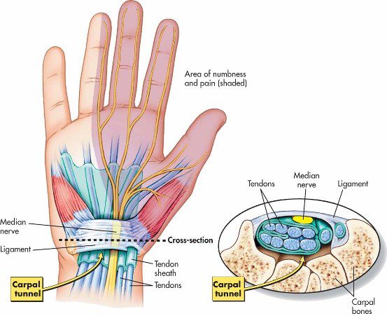
  • Tingling, numbness or burning sensation of the thumb, index, middle and ¾ of ring fingers of the hand
  • Electric shock like radiating pain through the hand into thumb, index, middle and ¾ of ring finger
  • Weakened grip, loss of dexterity and fine movements such as picking up a hair pin, buttoning clothes.
  • Hypersensitivity or in other cases lessened sensation of hand to pressure, heat or cold temperatures
  • Swollen wrist

Let’s take a closer look at the anatomy!

As its name suggests – a group of small bones aka carpal bones form a tunnel like passageway in the wrist (palmar view). This unique architectural design allows for the tendons of the forearm muscles and the all-important median nerve to pass through the narrow tunnel through the wrist and into the hand and fingers, supplying sensation and motor function.

Causes

Common causes and risk factors that increase the likelihood of carpal tunnel syndrome include:

  • Repetitive wrist & hands movements – during work related tasks or leisure activities may irritate the tendons in the wrist, resulting in inflammation that irritates the nerve.
  • Wrist or hand injury – recurring sprains, swelling and reduced wrist movements reduces the space in the carpal tunnel
  • Pregnancy and menopause – hormonal changes can increase fluid retention in body increasing pressure in the carpal tunnel compressing the median nerve
  • Genetic history – petite
  • Medical conditions (rheumatoid arthritis, diabetes, hyperthyroidism)

Interesting facts about carpal tunnel syndrome

  • Women are 3 times more susceptible to develop carpal tunnel syndrome than men. This can be due to hormonal changes during pregnancy or menopause and also because women tend to have smaller carpal tunnels.
  • Not all fingers are affected. Median nerve supplies movement and sensation in the thumb, all fingers except the little finger.
  • Computers/keyboard are not the only reasons to blame – repetitive nature of any work related or leisure word increases risk of developing carpal tunnel syndrome

Diagnosis

Carpal tunnel syndrome is fairly easily diagnosed by your physiotherapy, doctor or a hand therapist.

Your health practitioner will gather information on your general health, history and nature of your symptoms. They will then carefully conduct a thorough clinical assessment to assess the movements of your hand and wrist, strength and use a collection of tests in effort diagnose your symptoms. In some cases, your therapist may examine your neck, shoulders and arms to rule out other potential causes.

You may often hear the physiotherapist or hand therapist mention that they want to conduct a functional assessment – A functional assessment is activity specific, where the therapist will watch you perform the activity that aggravates your symptoms the fastest. For example, if using a keyboard is generally when you feel your symptoms start – the therapist may observe you performing the very task to examine your overall posture.

Referral to scans or nerve conduction tests may be arranged by your doctor or therapist depending on the severity or complexity of your symptoms.

Scans

Referral to scans or nerve conduction tests may be arranged by your doctor or therapist depending on the severity or complexity of your symptoms.

  • Xray – provides key information on bone health, when dealing with a potential injury, or arthritis
  • Ultrasound – can examine potential soft tissue injury or inflammation compressing the median nerve
  • MRI – this advanced imaging provides in depth review of your wrist and hand. Usually arranged by your doctor or a specialist
  • Nerve conduction study – studies the electrical activity of the median nerve. This test will help you doctor examine the severity of your problem.

Treatment

In most cases, carpal tunnel syndrome will progressively worsen overtime. So, the key is early intervention!

Conservative management

Mild symptoms can be easily managed with a conservative approach.

  • Wearing splints or braces – keeps your wrist straight to prevent repetitive use of hands, thus reducing pressure or inflammation in the carpal tunnel.
  • Non-steroidal anti-inflammatory medications – such as celecoxib and ibuprofen as prescribed by your doctor may decompress the median nerve by reducing the inflammation in your body and wrist.
  • Activity modification: your physiotherapist will play an important role in providing you with advice around to modifying your activities to reduce your symptoms. They will also prescribe you with effective stretches and exercises to help manage your symptoms while safely aiding your recovery.
  • Steroid injections: your physiotherapist or doctor may recommend a ‘cortisone’, also known as a ‘corticosteroid’ injection to control your symptoms. It contains an anti-inflammatory substance that is injected into your carpal tunnel. The effects of the steroid injection may be temporary and can vary person to person depending on many factors (cause of symptoms, stage of your condition).

In mild to moderate cases, the effects of injection may last between 3-6months.

Surgical intervention

If non-surgical approaches have failed to relieve your symptoms, surgery may be required.

By this stage you would have consulted an orthopaedic surgeon. Your surgeon will thoroughly examine your overall health, symptoms, results from the scans and the nerve conduction study to help you decide on the best treatment approach.

If you decide to undergo surgery – the surgical procedure your surgeon will perform is called ‘carpal tunnel release’.

Recovery and outcomes

  • After your surgery you may be given a splint or a brace for a period of time specified by your surgeon. While in the splint or brace you will be encouraged to move your fingers to prevent stiffness and swelling.
  • Expect to experience minor pain, stiffness and swelling for a couple of weeks to months after your surgery. Pain medications provided by your surgeon must be taken as prescribed.
  • You may be encouraged to see your physiotherapist, who will work closely with your surgeon to help meet post-operative outcomes.
  • You will have regular 6-8 weekly follow ups with your surgeon as required to assess your healing and discuss gradual return to light activities and return to work.
  • If you have underlying medical conditions such as arthritis, except that your recovery may be slower than otherwise expected. It is important that you follow post-operative protocols your surgeon, doctor and physiotherapist recommend.
Arthritis

ARE YOU IMPACTED BY ARTHRITIS ON A DAILY BASIS?

What is Osteoarthritis?  

Osteoarthritis is a condition that causes pain, discomfort and stiffness at our joints. It is the most common form of arthritis worldwide.

The intensity of the pain is different for each individual person. Osteoarthritis can occur at any joint, however it most commonly impacts the hips, knees and hands.


Signs/Symptoms

The symptoms of osteoarthritis usually start gradually and increase over time. You may experience pain, tenderness and clicking/crackling sounds with movement.

Swelling: Commonly occurs around the affected joints and soft tissues.

Pain: Joint pain usually increases during/after activity or movement. It can also increase due to inactivity.

Stiffness: Can be present first thing in the morning or after activity.

Reduced range of motion: You may be unable to move the joint freely through the normal range.

Causes:

In normal day to day life we are constantly loading our joints causing low level damage. The majority of the time our bodies heal naturally and you never experience any symptoms.

Osteoarthritis occurs when the cartilage that cushions our joints breaks down over time. This can lead to bony growth within our joints and inflammation. Eventually, our connective tissues that support our joints deteriorate.

Risk factors for developing Osteoarthritis:

  • Previous joint injury- Sporting or old injuries
  • Age Increased risk as you get older.
  • Sex – More common in females
  • Family history- There is some evidence of family/genetic links.
  • Obesity- Being overweight increases the stresses and strains on our joints.

Diagnosis

Clinical examination of the joint: assessing for signs of swelling, tenderness and/or loss of motion. Gathering information on the injury history and symptoms.

X-ray: Cartilage loss does not appear on X-ray imaging. However a narrowing of space between the bones can be seen. Bony spurs around the joint can also be identified.

MRI: Magnetic resonance imaging: Provides detailed images of soft tissue and bony structures. For the majority of cases it is not used to diagnose osteoarthritis but can help in more complex cases.

Treatment options: Conservative management

Osteoarthritis requires long-term management. Although there is no cure for osteoarthritis, treatments can reduce pain and get you moving better.

Mild symptoms/early stages:

  • low impact exercises – walking, swimming, cycling, strength training
  • reducing overall body weight (if overweight)
  • wearing supportive footwear
  • using aids/walking devices to reduce overall joint loading

Medication:

If your symptoms are more severe and you are finding the pain difficult to manage, speak to your GP or pharmacist about painkillers or anti-inflammatory drugs.

Physiotherapy:

  • Detailed assessment of your individual needs
  • Structured and progressive rehabilitation plan
  • Advice on self management

Non-conservative management:

If symptoms are more severe and you are not getting relief from the above options, you may benefit from procedures such as:

Steroid injection

An injection may be indicated to relieve persistent pain, following a trial of conservative management.

Surgery

Joint replacement surgery is an option in more severe cases, again following a trial of conservative management. Note: there is rehabilitation required before and after surgery. It is not a quick fix option.

How we can help

  • Provide a detailed assessment and advice on your current symptoms and best treatment options available.
  • Prescribe you a structured plan
  • Onward referral to a specialist or further imaging if indicated