fbpx
Your first step to recovery

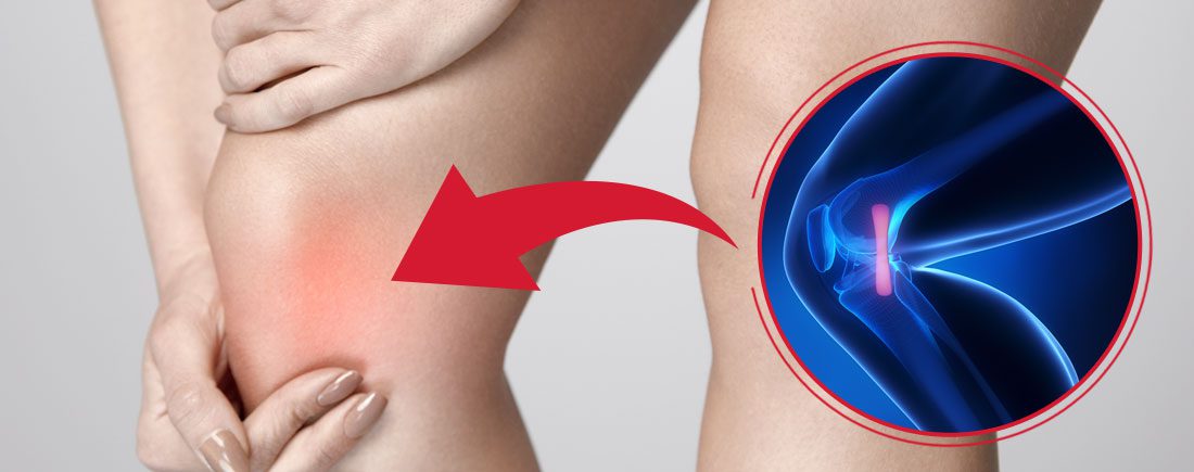
Managing your Medial Knee Pain: MCL injuries

What is it?

Although your knee has free movement going forwards and backwards, its’ sideward movements are restricted by the robust collateral ligaments on either sides of your knee. The medial collateral ligament (MCL) is situated on the inner part of your knee, but on the outside of your joint. The MCL connects the top of your shinbone (tibia) to the bottom of your femur (thighbone). It helps hold your bones together, provides stability and prevents your knee from bending sideways away from your body.

Injuries to the MCL are from the result of a direct blow to the outer part of your knee- and is most commonly seen in contact sports such as football and soccer. These injures may either over-stretch or cause a tear in the ligament. Whilst surgery may be needed in some severe cases, it is not always the go-to form of management.

Read on to know how physiotherapy can help manage your MCL related-knee pain.


Mechanism of Injury

Injury to the MCL typically occurs when a force drives the lower leg in a sideward direction away from your upper leg and body. Awkwardly landing from a height, twisting of your knee with your foot fixated to the ground, or from a direct blow to the outer part of your knee- most commonly seen in contact sports, are frequent causes of injury to the MCL.


Grading of MCL Injuries

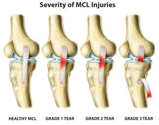
MCL injuries are often graded using the system below:

Grade 1: Regarded as a minor injury- means that the MCL has been overstretched but not torn

Grade 2: Regarded as a moderate injury- means that there is a partial tear in MCL, and presents with some degree of instability in the knee

Grade 3: Regarded as a severe injury- means that the MCL has completely ruptured/torn, and presents with noticeable joint instability


Often 3 MCL injuries are associated with concurrent medial meniscus and ACL ligament damage, which may need surgical intervention. But, the good news is that most MCL injuries may be treated well with conservative physiotherapy management. It usually takes between 2-8 weeks for Grade 1 and 2 MCL injuries to heal, and a graduated rehabilitation programme is highly commended for prevention of future injury.


Signs and Symptoms

Because injury to the MCL may present with similar symptoms as with other knee injuries such as ACL damage, it is vital to have a medical professional such as your physiotherapist evaluate your injury.

Common symptoms of an MCL injury may include:

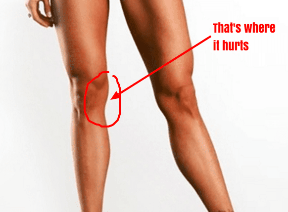
  • Tenderness and pain along in the inner part of your knee
  • Swelling in the knee
  • Experience catching and locking sensations in the knee joint
  • A ‘pop’ sound at the time of injury
  • Actual or feeling of giving way of the knee (often indicate grade 2 or 3 injury)
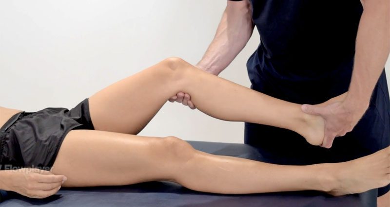


Diagnosis

Your physiotherapist will discuss your injury and its presenting symptoms, past medical history (including a history of any prior knee injuries) and will also undertake a thorough physical examination. During the physical examination, your physiotherapist will assess the structures of your injured knee and compare them to the non-injured side. The range of motion, strength and stability of your knee will be assessed. You may be referred on for imaging such as X-rays and Ultrasounds to help aid the diagnosis. For more severe MCL injuries, and if your symptoms do not resolve with conservative physiotherapy management, you may be referred onto a specialist who may consider referring you for an MRI to get a deeper look at your knee.


Management

The management options for MCL injuries will be dependent on the severity of the injury. In the initial stages of injury, management is focused on controlling swelling and pain, whilst allowing your body to initiate healing processes via inflammation. This is typically achieved through the P.O.L.I.C.E. principles (Protect, Optimal Loading, Ice, Compression and Elevation).

Over the counter medication such as ibuprofen and paracetamol may be taken to reduce pain. Other stronger painkillers and NSAIDs may be prescribed by your doctor to help reduce swelling and inflammation as well.

After assessing your knee, your physiotherapist will frame a rehabilitation programme with exercises tailored to your needs. The purpose of physiotherapy is to help restore your knee’s range of motion, stability and strength, which in turn will then allow you to safely return to your usual day-to-day and sporting activities as soon as possible.

Management of most MCL injuries usually only involves knee bracing and physiotherapy treatment. However, in some cases, surgery may be recommended. Particularly if there is damage to more than one ligament or structure in your knee or if you continue to experience instability in spite of physiotherapy.

Ankle injuries – Your first step to recovery

Common? Oh Yes!

The ankle is the most commonly injured joint in sport. This does not exclude other people such as active hikers, beach goers and even your average Sunday stroller. Good news though – your ankle injury is highly likely to be uncomplicated.

It is still vital that your ankle is examined, evaluated and treated early. This will ensure a swift return to activity and prevent further complications.


The road to recovery

Planning for Resiliency and the Road to Recovery

Your clinician will ask you some questions related to how you injured your ankle, pain, instability and any past episodes of injury. The earlier you get your ankle checked, the sooner your recovery will begin.

Keeping a mental note of things like initial pain, swelling, ability to walk and balance will go a long way in assisting your clinician to making an accurate diagnosis.


Investigations

Ankle x-rays

In most cases, initial X – rays are done to rule out broken bones.

Ultrasounds can be used to diagnose some ligament and tendon damage.

MRI is the best form of imaging but this does come at a higher cost and higher exposure to radiation. These are usually done after failed conservative treatment or in instances where pain remains high for longer periods.

A CT scan is helpful with complicated foot and ankle fractures. It will normally be ordered by a specialist surgeon who is planning for an operation.


What to look out for

Ankle sprains:

This is normally a twisting injury that causes a stretch or tear of ligaments surrounding the ankle. Your health care professional will provide you with all the information and tools you need for recovery.

These heal relatively quickly when the outside border of the foot is affected and a little slower when the inside border of the ankle is affected.

You will normally feel pain on certain ankle movements, stiffness in the ankle and experience some swelling and bruising.

Ankle - Wikipedia


Ankle Fractures:

These normally present with swelling, bruising and pain initially – although not always. In some cases, it is too painful to put weight on the ankle.

They are usually best confirmed with X – ray and specialist referral.

Management may be surgical or non-surgical depending on the severity and site of the fracture.

Fractures generally take longer to recover compared to sprains.


What treatment to expect

Foot and Ankle Doctor | EmergeOrtho—Triangle Region

Acute phase:

Your healthcare professional will normally initiate techniques to minimise your pain and swelling with rest, ice, compression and elevation.

Analgesia and anti – inflammatory medication may also be used.

Strapping may be used for stability at this stage and can be done by your physiotherapist.

You will also be encouraged to increase movement and begin strengthening.


Rehabilitation phase:

Balance and proprioceptive exercises will be given to you by your physiotherapist.

Strengthening will continue and running will start soon.

Once running in a linear motion pain free, you will progress to sport specific exercises.

Finally, you will return to sport or previous function such as trekking with a graded program.

Strapping may continue for up to 12 months after your injury in order to prevent re–injury.


What can you do on the day of the injury?

Rest by reducing time spent walking or standing. This will help the ankle to heal.

Ice the ankle for up to 20 minutes every couple of hours.

Compress the ankle with a firm bandage during the day and remove the bandage at night.

Elevate the leg.

Attempt circulatory exercises such as ankle circles and foot pumps (About 10 – 30 repetitions every couple of hours).

Contact your health professional or physiotherapist in order to make appointment for assessment.

If you are unable to stand on your leg or have excruciating pain in the ankle, head on to the local emergency department for immediate investigation.


Remember, your injury will heal and you will recover!


To find your nearest Physio Fusion clinic and book an appointment call 09 6266186 or visit our website https://physiofusion.co.nz

The myth about foot pronation (flat foot)

Myth: Foot pronation(flat foot) is the enemy.

Quest - Article - Surgery Sometimes, Bracing Often, Caution Always |  Muscular Dystrophy Association

In the foot, pronation should occur naturally when the foot comes into contact with the ground. Pronation will appear as the foot rolling inward and the arch flattening.

What are the benefits?

  • Dissipates the force that the foot receives from the ground

  • Allows the foot to become a stable and mobile adaptor to enhance movement opportunity

  • Loads the muscles of the extensor chain (calf, quads, glutes) to convert ground reaction forces into forward momentum so we can propel efficiently.

So why have I been told this is bad?

So as you are now aware, pronation is very normal and a critical movement to ensure we move and propel ourselves forward efficiently.

What you may have heard someone say to you is that you ‘overpronate’?

Firstly, overpronation is subjective and not as black and white as it is sometimes made out to be. Overpronation has be defined as: ‘a foot that rolls inward toward the arch excessively’.

Foot Pronation: Underpronation & overpronation explained- The Foot Clinic

What we must understand is that a pronation can only happen when the foot has a stable tripod on the ground. This means that the calcaneus (heel bone), 1st metatarsal (big toe knuckle), 5th metatarsal (little toe knuckle) must all remain in contact when the foot rolls inwards and the arch flattens.

The Foot Tripod - Fix Flat Feet

So, If you have been told you are ‘overpronated’ , it is most likely that your whole foot is ‘everting’ NOT ‘overpronating’.

What is Eversion?

Eversion can be defined as: ‘the process of turning inside-out’.

In pronation your heel must naturally ‘evert’ (sole of the heel will move away from the midline of the body) NOT your whole foot.

If your ‘whole foot’ everts (turns out) you will no longer have a stable foot tripod as the 5th metatarsal (little toe) will lose contact with the ground.

The key to ensuring this does not happen is to provide an environment for the bones of the midfoot (middle of the foot) and forefoot (toes) to experience the opposite motion to that of the heel. This will mean that the foot has an opportunity to truly pronate with a tripod on the ground.


So how can you help me do that?

  • At Physio fusion we can help you to bring your own body into alignment and create an environment in which the healing can begin

  • Foot strengthening exercises

  • Footwear advice

  • Referral to other healthcare specialists for further assistance (e.g. podiatrists)

To find your nearest Physio Fusion clinic and book an appointment call 09 6266186 or visit our website https://physiofusion.co.nz

Disorders of the Achilles Tendon

Basic Anatomy

The Achilles tendon is the largest tendon in the human-body. It is a band of tissue that connects your calf muscles to your heel bone (calcaneus). This tendon primarily facilitates general mobility such as walking, running, climbing stairs, jumping, and standing on your tip toes, by helping to raise the heel off the ground.


Common Achilles Pathology

Achilles tendinitis and tendinosis are two common disorders and are typically classified as overuse injuries.

Achilles tendonitis involves inflammation of the Achilles tendon. Inflammation is the body’s natural response to injury or disease, and often causes swelling, pain, or irritation. This inflammation is typically short-lived. Over time, if this is left resolved, the condition may progress to degeneration of the tendon- Achilles tendinosis, in which case, the tendon loses its organized structure and is likely to develop microscopic tears.

There are two types of Achilles tendonitis and it is based on which part of the tendon is inflamed:

  • Insertional Achilles tendonitis affects the lower portion of your tendon where it attaches to your heel bone.
  • Non-insertional Achilles tendonitis involves fibres in the middle portion of the tendon and tends to affect younger people who are active.

In both non-insertional and insertional Achilles tendinitis, damaged tendon fibres may also calcify (harden) and often bone spurs (extra bone growth) develop with insertional Achilles tendinitis. Achilles tendonitis may also increase your risk of sustaining an Achilles tendon rupture (tear).

Causes

Typically referred to as “overuse” conditions, Achilles tendonitis and tendinosis are often caused by the sudden increase in repetitive activity involving the Achilles tendon. This can put too much stress on the tendon too quickly, that can then lead to micro-injury of the tendon fibres. Because of this ongoing stress on the Achilles, the body is not able to repair the injured tissue. The structure of this tendon is then modified, resulting in continued pain and other symptoms. The Achilles tendon also has poor blood supply that makes it more susceptible to injury and may make recovery from injury slow.

Common factors that may lead to the development of disorders of the Achilles tendon include:

  • Weak and/or tight calf muscles
  • Rapidly increasing the amount or intensity of exercise within a short span of time
  • Hill climbing or stair climbing exercises
  • Presence of bony spurs in the back of your heel
  • Changes in footwear – especially changing from wearing high-heeled shoes to flat shoes
  • Wearing poor fitting, inappropriate, or worn out shoes during sporting activities
  • Exercising without adequate warm-ups and stretching
  • A sudden sharp movement which causes the calf muscles to contract and the stress on the Achilles tendon to be increased. This can cause the tendon fibres to tear.
  • Excessive mobility
  • Poor feet positioning and biomechanics (excessive pronation and flattening of the arches of the foot)


Symptoms

Achilles tendon pain: Causes. when to see a doctor, and treatment


Common symptoms include:

  • Pain and stiffness along the Achilles tendon especially first thing in the morning
  • Pain along the tendon or back of the heel that worsens with activity
  • Severe pain the day after exercising
  • Visible thickening of the tendon
  • Tenderness to touch
  • Bone spur
  • Swelling that is present all the time and gets worse throughout the day with activity

If you have experienced a sudden “pop” in the back of your calf or heel, you may have torn your Achilles tendon. Please seek urgent medical attention if you think you may have torn your tendon.

Diagnosis

If Achilles tendonitis or tendinosis is suspected, please deter from any activity or exercise which causes the pain. It is advisable to see your doctor or physiotherapist as soon as possible so that an accurate diagnosis may be made and appropriate treatment recommended.

You will be asked about the nature and duration of your symptoms and the medical professional assessing you will have a look at your foot and ankle. Ultrasound scanning may be used to evaluate the damage to the tendon and/or surrounding structures.

An MRI may be recommended if symptoms persist. X-rays may also be taken to rule out other disorders which may cause symptoms like Achilles tendonitis and tendinosis.

Achilles Tendonitis - Ankle - Conditions - Musculoskeletal - What We Treat  - Physio.co.ukHow to Treat Achilles Tendinopathy with Physical Therapy -  prohealthcareproducts.com

Treatment

Treatment will depend on the nature, severity, and length of the injury. Generally speaking, the longer the symptoms are present before treatment commences, the longer the timeframe until full recovery is attained.  Full recovery may take between three and nine months.

Initial treatment options in the early stages may include:

  • Rest – to avoid further injury to the area
  • Ice – to reduce inflammation
  • Elevation – to reduce swelling
  • Non-steroidal anti-inflammatory drugs to reduce pain and inflammation.

 How physiotherapy can help:

Physiotherapy typically focuses on two main areas: treatment and rehabilitation. Treatment may entail massage, shockwave therapy, acupuncture, gait re-education, and gentle stretching, whereas, rehabilitation predominantly entails strengthening of the Achilles and surrounding musculature.

Strengthening of the muscles surrounding the Achilles tendon facilitates healing in the tendon itself. Strengthening is attained through the utilization of specific exercises, that will be taught by your physiotherapist. It is common for the rehabilitation programme to take up to three months.


Exercises