fbpx
Your first step to recovery

Managing Your Osteoarthritis

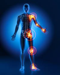
Osteoarthritis impacts millions of people worldwide and is typically known as the most common form of arthritis. It is associated with the wear and tear of the protective cartilage which cushions the ends of your bones in your joints over time. Though this condition may cause damage to any joint in the body, osteoarthritis primarily impacts the joints in your spine, hands, hips, and knees.


Causes and Risk factors

Over time, the gradual deterioration of the cartilage which cushions the ends of your bones in your joints causes arthritis. Cartilage is a solid slippery tissue which allows almost frictionless joint movement. As the cartilage wears down, bone will eventually rub on bone.

This condition is typically characterized as a wear and tear degenerative disorder. However, in addition to the breakdown of the cartilage, it also impacts the joint as a whole. Osteoarthritis triggers alterations in the bone and damages connective tissue which holds the joint together and attaches your muscles to your bones. Inflammation of the lining of the joint is also triggered.

Factors which may put you at higher risk of developing osteoarthritis include but are not limited to:

  • Your age- the risk increases with getting older
  • Gender- though unclear why, but women are more perceptible to developing osteoarthritis
  • Bony deformities- those with abnormal joints or defective cartilage
  • Sustaining bony or joint injuries like those which take place during sport or from an accident.
  • The risk increases with obesity- the more you weigh, the greater your risk, as it adds more stress to your weight-bearing joints (particularly hips and knees)
  • Your occupation or a sport that you play which puts repetitive and excessive stress/loading on the joints, can eventually lead to the development of osteoarthritis.
  • Certain co-morbidities such as diabetes


Common symptoms

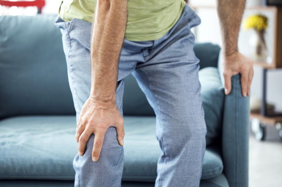
Below are some common examples of symptoms you may experience with arthritis. These may develop and worsen gradually over time

  • Pain: Your joints may hurt before and/or after undertaking an activity
  • Loss of joint range of motion– loss of overall joint flexibility and movement
  • Tenderness felt on applying light pressure to the joint
  • Joint stiffness that is most noticeable on waking up first thing in the morning or after a prolonged period of inactivity
  • Noticeable changes in joint pain with changes in the weather- particularly colder weathers
  • Sensations of grating and grinding// sounds of clicking and popping (crepitus) when you use the joint
  • You may notice swelling and redness around the joint, which may be triggered by soft tissue inflammation
  • Bony spurs that feel like hard bumps may develop around the impacted joint


How will I be diagnosed?

Osteoarthritis is typically diagnosed based on your medical and occupation history and a physical examination undertaken by your doctor. During the physical examination, your doctor will assess your affected joint(s) for swelling, tenderness, redness, and stiffness. X-rays may be recommended to reveal cartilage loss (the narrowing of the space between the bones of your joints), changes in bone, and bony spurs around the joint. Blood tests may be used to rule out other causes of joint pains like rheumatoid arthritis. Joint fluid analyses may also be used to test for inflammation to ascertain if the pain is triggered by an infection or gout instead of osteoarthritis.


Management

Though there isn’t a cure for osteoarthritis, various treatments which can help relieve symptoms of pain and disability are available.

Lifestyle modifications: Changes to your daily life may protect your joints and slow the progression of osteoarthritis.  Minimising activities which exacerbate your symptoms such as climbing stairs, squatting. Swapping high-impact activities like running and jogging to lower-impact activities such as cycling or hydrotherapy will decrease the stress on your joints. Weight-loss reduces the stress and loading on your joints, which then results in less pain with increased function.

Assistive aids: Using assistive aids like a stick/cane, wearing proper shoes w orthotics and supportive braces/sleeves may improve your stability and support your functional capabilities.

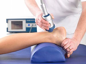
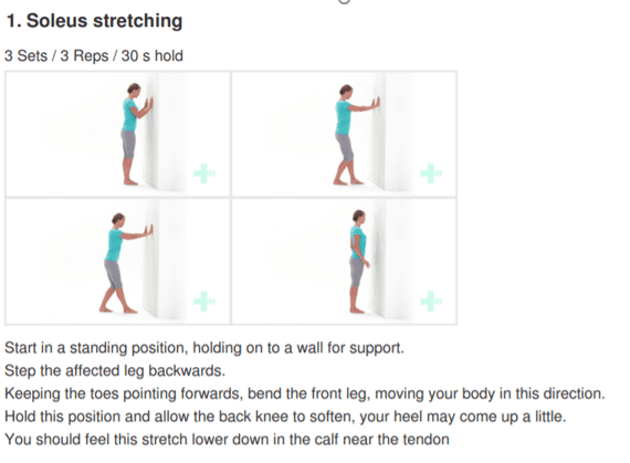
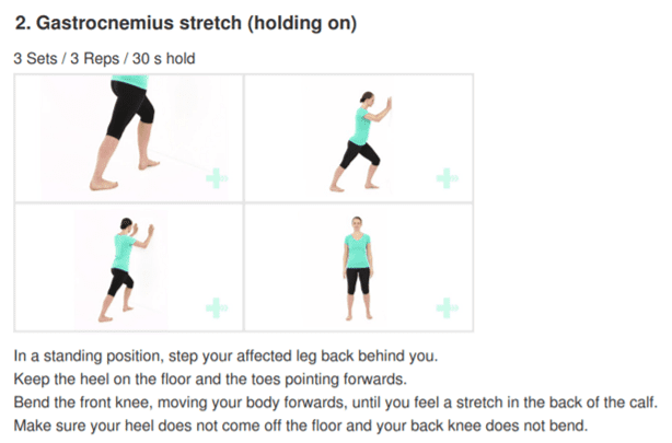
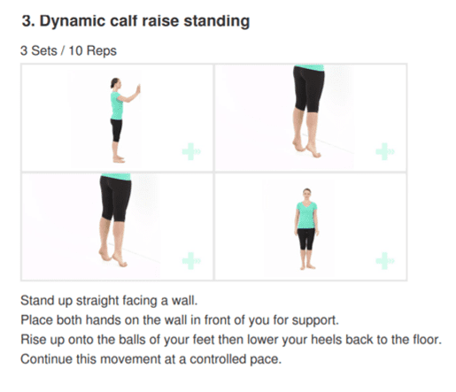
Physiotherapy: Targeted exercises may help improve your flexibility as well as build strength in your muscles. Your physiotherapist will develop a personalised active rehabilitation program which is safe and will meet your requirements and lifestyles.

Medications: Various kinds of medication (such as paracetamol and NSAIDs) maybe helpful in treating and controlling the symptoms of osteoarthritis. As everyone responds differently to medications, your doctor will prescribe medicines (type and dosage), which is safe and will work best for you.

Cortisones: Strong anti-inflammatory agents which is injected into the affected joint to give pain relieve and decrease inflammation for a short period of time. Due to potential side-effects, it may be recommended to restrict the number of injections to 2-3 per year.

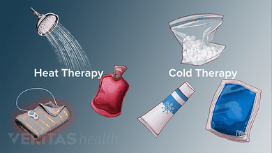
Other: Heat and ice applications, self-massaging with pain-relieving creams/ointments and/or wearing elastic supports may provide some relief from your pain and give you support.

Surgery: Surgery may be recommended if there is considerable degeneration in your joints and/or if your osteoarthritic pain causes disability that is not relieved with conservative management. Your doctor or specialist will discuss your options with you.

Carpal Tunnel Syndrome – What is it?

Have you been experiencing pain, pins and needles or numbness in your wrist and hands, especially after using the keyboard, chopping up a few veges, reading a book, using your mobile phone or with driving?

If you answered yes – then you are most likely to have Carpal tunnel syndrome.

What is Carpal Tunnel Syndrome?

Carpal tunnel syndrome is the most common condition in the arm. It is caused by compression of one of the three major nerves in the forearm – the median nerve, which travels through the wrist into the hand and fingers. Entrapment of the median nerve usually due to inflammation, occurs in the wrist commonly resulting in tingling of the wrist and hand (in some cases forearm), numbness, pain and weakness of the hand.

Signs and Symptoms

Often unrelated to a specific incident or an injury, symptoms of carpal tunnel syndrome usually develop gradually overtime. Symptoms may be worse in the morning and night. Many people find that the frequency and duration of symptoms increase as the conditions worsen.

Signs and symptoms may include:

  • Tingling, numbness or burning sensation of the thumb, index, middle and ¾ of ring fingers of the hand
  • Electric shock like radiating pain through the hand into thumb, index, middle and ¾ of ring finger
  • Weakened grip, loss of dexterity and fine movements such as picking up a hair pin, buttoning clothes.
  • Hypersensitivity or in other cases lessened sensation of hand to pressure, heat or cold temperatures
  • Swollen wrist

Let’s take a closer look at the anatomy!

As its name suggests – a group of small bones aka carpal bones form a tunnel like passageway in the wrist (palmar view). This unique architectural design allows for the tendons of the forearm muscles and the all-important median nerve to pass through the narrow tunnel through the wrist and into the hand and fingers, supplying sensation and motor function.

Causes

Common causes and risk factors that increase the likelihood of carpal tunnel syndrome include:

  • Repetitive wrist & hands movements – during work related tasks or leisure activities may irritate the tendons in the wrist, resulting in inflammation that irritates the nerve.
  • Wrist or hand injury – recurring sprains, swelling and reduced wrist movements reduces the space in the carpal tunnel
  • Pregnancy and menopause – hormonal changes can increase fluid retention in body increasing pressure in the carpal tunnel compressing the median nerve
  • Genetic history – petite
  • Medical conditions (rheumatoid arthritis, diabetes, hyperthyroidism)

Interesting facts about carpal tunnel syndrome

  • Women are 3 times more susceptible to develop carpal tunnel syndrome than men. This can be due to hormonal changes during pregnancy or menopause and also because women tend to have smaller carpal tunnels.
  • Not all fingers are affected. Median nerve supplies movement and sensation in the thumb, all fingers except the little finger.
  • Computers/keyboard are not the only reasons to blame – repetitive nature of any work related or leisure word increases risk of developing carpal tunnel syndrome

Diagnosis

Carpal tunnel syndrome is fairly easily diagnosed by your physiotherapy, doctor or a hand therapist.

Your health practitioner will gather information on your general health, history and nature of your symptoms. They will then carefully conduct a thorough clinical assessment to assess the movements of your hand and wrist, strength and use a collection of tests in effort diagnose your symptoms. In some cases, your therapist may examine your neck, shoulders and arms to rule out other potential causes.

You may often hear the physiotherapist or hand therapist mention that they want to conduct a functional assessment – A functional assessment is activity specific, where the therapist will watch you perform the activity that aggravates your symptoms the fastest. For example, if using a keyboard is generally when you feel your symptoms start – the therapist may observe you performing the very task to examine your overall posture.

Referral to scans or nerve conduction tests may be arranged by your doctor or therapist depending on the severity or complexity of your symptoms.

Scans

Referral to scans or nerve conduction tests may be arranged by your doctor or therapist depending on the severity or complexity of your symptoms.

  • Xray – provides key information on bone health, when dealing with a potential injury, or arthritis
  • Ultrasound – can examine potential soft tissue injury or inflammation compressing the median nerve
  • MRI – this advanced imaging provides in depth review of your wrist and hand. Usually arranged by your doctor or a specialist
  • Nerve conduction study – studies the electrical activity of the median nerve. This test will help you doctor examine the severity of your problem.

Treatment

In most cases, carpal tunnel syndrome will progressively worsen overtime. So, the key is early intervention!

Conservative management

Mild symptoms can be easily managed with a conservative approach.

  • Wearing splints or braces – keeps your wrist straight to prevent repetitive use of hands, thus reducing pressure or inflammation in the carpal tunnel.
  • Non-steroidal anti-inflammatory medications – such as celecoxib and ibuprofen as prescribed by your doctor may decompress the median nerve by reducing the inflammation in your body and wrist.
  • Activity modification: your physiotherapist will play an important role in providing you with advice around to modifying your activities to reduce your symptoms. They will also prescribe you with effective stretches and exercises to help manage your symptoms while safely aiding your recovery.
  • Steroid injections: your physiotherapist or doctor may recommend a ‘cortisone’, also known as a ‘corticosteroid’ injection to control your symptoms. It contains an anti-inflammatory substance that is injected into your carpal tunnel. The effects of the steroid injection may be temporary and can vary person to person depending on many factors (cause of symptoms, stage of your condition).

In mild to moderate cases, the effects of injection may last between 3-6months.

Surgical intervention

If non-surgical approaches have failed to relieve your symptoms, surgery may be required.

By this stage you would have consulted an orthopaedic surgeon. Your surgeon will thoroughly examine your overall health, symptoms, results from the scans and the nerve conduction study to help you decide on the best treatment approach.

If you decide to undergo surgery – the surgical procedure your surgeon will perform is called ‘carpal tunnel release’.

Recovery and outcomes

  • After your surgery you may be given a splint or a brace for a period of time specified by your surgeon. While in the splint or brace you will be encouraged to move your fingers to prevent stiffness and swelling.
  • Expect to experience minor pain, stiffness and swelling for a couple of weeks to months after your surgery. Pain medications provided by your surgeon must be taken as prescribed.
  • You may be encouraged to see your physiotherapist, who will work closely with your surgeon to help meet post-operative outcomes.
  • You will have regular 6-8 weekly follow ups with your surgeon as required to assess your healing and discuss gradual return to light activities and return to work.
  • If you have underlying medical conditions such as arthritis, except that your recovery may be slower than otherwise expected. It is important that you follow post-operative protocols your surgeon, doctor and physiotherapist recommend.

How to stay Healthy and Sane during Lockdown


The restrictions and change brought by the outbreak of COVID-19 has resulted in a great deal of control being taken from our hands; this has been anxiety provoking for many of us. Nevertheless, it’s important to re-evaluate, acknowledge and place focus upon the matters that we DO have control over so that we can gain our personal power back!


Lockdown Productivity Tips


Check in with yourself: how is your body and mind feeling. Embrace your emotions and give yourself permission to feel the way toy do.

:large_blue_diamond: Stay connected: Social connection is inevitably limited at the moment but catching up with people via text or facetime will help prevent feelings of isolation.

:large_blue_diamond: Maintain some form of routine: Maintaining a routine helps provide some structure do days which often all seem to merge into one.

:large_blue_diamond: Get fresh air where possible: Daily fresh air can provide an easy change of scenery when we are stuck at home most of the day.

:large_blue_diamond: Gentle exercise is a MUST!

:large_blue_diamond: Stay Hydrated: Drinking enough water is important to keep your body hydrated and makes sure your body functions properly.

:large_blue_diamond: Eat well- You’d be surprised how your diet can affect how you feel. Gut health in particular is linked to mental health.

:large_blue_diamond: Get to that “thing” you’ve been delaying for months

:large_blue_diamond: Pick up a good book

:large_blue_diamond: Learn new habits or rediscover old ones

:sparkles: These may seem like simple strategies but sometimes it’s the simple things that are most effective :sparkles:

“One day this will all be over and we will be grateful for life in ways we never felt possible”

The gratitude we will have for the things we once took for granted will be unmeasurable- getting on a plane, an impromptu visit to the cinema, a shopping spree, going to the gym, even meeting a friend for lunch at a café. Keep going, nothing lasts forever and we have so much to look forward to. In the mean time take each day as it comes, be kind, support those who are struggling and keep going! You are stronger and more resilient than you know!

Lifting Mechanics

IS IT DANGEROUS TO LIFT WITH A BENT BACK?

One common belief about lifting is that rounding your back when lifting an object is considered dangerous while lifting with a straight back is considered safe…


However, there is a lot of misinformation circulating around lifting mechanics and what is deemed “good technique” versus “bad technique.”

Here are some key myths and misinformation that you may have heard:


A ROUNDED BACK WHILE LIFTING OVER STRESSES THE BACK MUSCLES AND LIGAMENTS

  • Research has shown that regardless of lifting position, whether you’re stooped, squatting or weightlifting, your back has to produce the same amount of force. Statistically speaking, it’s not significantly different.


FLEXING THE SPINE WHILE LIFTING INCREASES THE LOAD ON THE LUMBAR DISCS

  • Under heavy loads, discs are unlikely to fail unless >95% flexion is achieved (which is near impossible)

  • With low loads, the disc is unlikely to fail unless you do thousands of repetitions continuously

  • Your body is an amazing adapter, this includes discs! Your discs can adapt and become more and more able to handle loads when stressed appropriately


THESE COMPOUND TO CAUSE INJURIES AND PAIN TO THE BACK MUSCLES AND DISCS

  • Resistance training has demonstrated through countless studies the ability to increase bone mineral density (BMD) of the lumbar spine. BMD is actually positively associated with the strength of the spinal discs and ligaments at that level. With appropriate loading and training, disc, ligament, bone and muscles are going to adapt favourably


A NEUTRAL SPINE IS SAFER, STRONGER, MORE EFFICIENT AND BETTER TO LIFT WITH

  • There is no significant difference between activities that encourage more spinal flexion and one’s that do not in the long term

  • Lifting with lumbar flexion is not a risk factor for low back pain

  • Research has shown lifting with a bent back is more metabolically and neuromuscularly efficient

  • When the spine is in extension during bent over activities, the hip is actually flexed to a greater degree – decreasing the ability for the glutes and hamstrings to create as much internal torque. Flexing the spine reduces this effect and reduces the moment arm for the hip extensors


STOP BACK PAIN & INJURIES BY LIFTING WITH A NEUTRAL SPINE

  • Your lumbar spine flexes every time your hip flexes! It is impossible to isolate one versus the other. It is also impossible to not flex while doing common movements

  • Extreme flexion however (>/=100%), may pose an increased risk under heavy loads, but not at light loads


You may still be wondering why you have back pain (stay tuned for our next blog!). Your pain may not be directly related to your lifting strategy.


Could you have pain with forward bending? Absolutely.

Is bending at the lumbar spine an increased risk for pain or injury? In the vast majority of situations, no.

If I have pain with forward bending, is it bad to temporarily limit doing so? Not at all.

Should I fear bending at the spine with or without pain or injury? No. Being fearful of flexing/moving your spine is actually a stronger predictor of disability and back pain.


Work on moving through your spine, after all it’s what it is designed to do! Choose comfortable movements and gain confidence over time, then build up your strength gradually with resistance training – try and not to push too fast, we want nice, healthy adaptation! Give your body time to adapt, back pain is not quick fix sometimes but you are resilient and with healthy, normal movements and some patience, you will be okay.

Disorders of the Achilles Tendon

Basic Anatomy

The Achilles tendon is the largest tendon in the human-body. It is a band of tissue that connects your calf muscles to your heel bone (calcaneus). This tendon primarily facilitates general mobility such as walking, running, climbing stairs, jumping, and standing on your tip toes, by helping to raise the heel off the ground.


Common Achilles Pathology

Achilles tendinitis and tendinosis are two common disorders and are typically classified as overuse injuries.

Achilles tendonitis involves inflammation of the Achilles tendon. Inflammation is the body’s natural response to injury or disease, and often causes swelling, pain, or irritation. This inflammation is typically short-lived. Over time, if this is left resolved, the condition may progress to degeneration of the tendon- Achilles tendinosis, in which case, the tendon loses its organized structure and is likely to develop microscopic tears.

There are two types of Achilles tendonitis and it is based on which part of the tendon is inflamed:

  • Insertional Achilles tendonitis affects the lower portion of your tendon where it attaches to your heel bone.
  • Non-insertional Achilles tendonitis involves fibres in the middle portion of the tendon and tends to affect younger people who are active.

In both non-insertional and insertional Achilles tendinitis, damaged tendon fibres may also calcify (harden) and often bone spurs (extra bone growth) develop with insertional Achilles tendinitis. Achilles tendonitis may also increase your risk of sustaining an Achilles tendon rupture (tear).

Causes

Typically referred to as “overuse” conditions, Achilles tendonitis and tendinosis are often caused by the sudden increase in repetitive activity involving the Achilles tendon. This can put too much stress on the tendon too quickly, that can then lead to micro-injury of the tendon fibres. Because of this ongoing stress on the Achilles, the body is not able to repair the injured tissue. The structure of this tendon is then modified, resulting in continued pain and other symptoms. The Achilles tendon also has poor blood supply that makes it more susceptible to injury and may make recovery from injury slow.

Common factors that may lead to the development of disorders of the Achilles tendon include:

  • Weak and/or tight calf muscles
  • Rapidly increasing the amount or intensity of exercise within a short span of time
  • Hill climbing or stair climbing exercises
  • Presence of bony spurs in the back of your heel
  • Changes in footwear – especially changing from wearing high-heeled shoes to flat shoes
  • Wearing poor fitting, inappropriate, or worn out shoes during sporting activities
  • Exercising without adequate warm-ups and stretching
  • A sudden sharp movement which causes the calf muscles to contract and the stress on the Achilles tendon to be increased. This can cause the tendon fibres to tear.
  • Excessive mobility
  • Poor feet positioning and biomechanics (excessive pronation and flattening of the arches of the foot)


Symptoms

Achilles tendon pain: Causes. when to see a doctor, and treatment


Common symptoms include:

  • Pain and stiffness along the Achilles tendon especially first thing in the morning
  • Pain along the tendon or back of the heel that worsens with activity
  • Severe pain the day after exercising
  • Visible thickening of the tendon
  • Tenderness to touch
  • Bone spur
  • Swelling that is present all the time and gets worse throughout the day with activity

If you have experienced a sudden “pop” in the back of your calf or heel, you may have torn your Achilles tendon. Please seek urgent medical attention if you think you may have torn your tendon.

Diagnosis

If Achilles tendonitis or tendinosis is suspected, please deter from any activity or exercise which causes the pain. It is advisable to see your doctor or physiotherapist as soon as possible so that an accurate diagnosis may be made and appropriate treatment recommended.

You will be asked about the nature and duration of your symptoms and the medical professional assessing you will have a look at your foot and ankle. Ultrasound scanning may be used to evaluate the damage to the tendon and/or surrounding structures.

An MRI may be recommended if symptoms persist. X-rays may also be taken to rule out other disorders which may cause symptoms like Achilles tendonitis and tendinosis.

Achilles Tendonitis - Ankle - Conditions - Musculoskeletal - What We Treat  - Physio.co.ukHow to Treat Achilles Tendinopathy with Physical Therapy -  prohealthcareproducts.com

Treatment

Treatment will depend on the nature, severity, and length of the injury. Generally speaking, the longer the symptoms are present before treatment commences, the longer the timeframe until full recovery is attained.  Full recovery may take between three and nine months.

Initial treatment options in the early stages may include:

  • Rest – to avoid further injury to the area
  • Ice – to reduce inflammation
  • Elevation – to reduce swelling
  • Non-steroidal anti-inflammatory drugs to reduce pain and inflammation.

 How physiotherapy can help:

Physiotherapy typically focuses on two main areas: treatment and rehabilitation. Treatment may entail massage, shockwave therapy, acupuncture, gait re-education, and gentle stretching, whereas, rehabilitation predominantly entails strengthening of the Achilles and surrounding musculature.

Strengthening of the muscles surrounding the Achilles tendon facilitates healing in the tendon itself. Strengthening is attained through the utilization of specific exercises, that will be taught by your physiotherapist. It is common for the rehabilitation programme to take up to three months.


Exercises