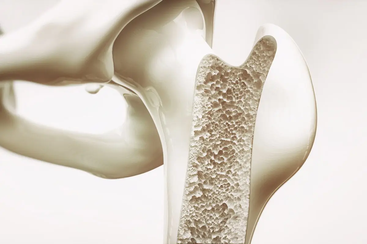
fbpx
Your first step to recovery

Osteoporosis


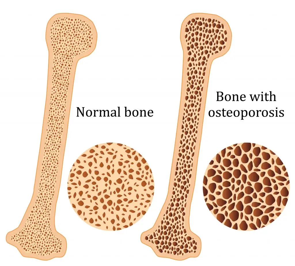
Osteoporosis is a condition which results in weak and brittle bones- to such degree that a fall or even mild stresses like coughing or bending over may result in a fracture. Bones are living tissues which are continually being broken down and replaced. However, your bones become osteoporotic when the formation of new bone does not keep up with the loss of old bone. This condition typically develops over time without any pain or other major symptoms, and is generally not diagnosed until you have sustained a fracture. The hip, pelvis, upper arm, spine and wrists are the most common structures affected by osteoporosis- related fractures.


How do you know if you have Osteoporosis?


Because there are no obvious early warning signs and symptoms, it is difficult to pre-diagnose osteoporosis. You may be unaware that you have this condition perhaps till you have one of the following:

  • Sustained a fracture from an incident more easily than you should have- like a simple fall or a bump
  • A decrease in the height of your spinal vertebrae over time
  • Change in posture – stooping or bending forwards
  • Back pain, due to a fractured or collapsed vertebra

Please see your doctor if you experience the following:

  • If you are over the age of 50 and have sustained a fracture
  • Sustained a spine, wrist, or hip for the first time
  • Sustained a fracture more easily than you should have (a simple fall or after a slight bump)


Risk factors

Key factors which may increase your risk of developing osteoporosis include:

  • Females- particularly post-menopausal Caucasian and Asian women
  • Over the age of 50
  • Excessive consumption of caffeine or alcohol
  • Smoking
  • Having a smaller or petite body frame
  • Poor physical activity levels and leading a very sedentary lifestyle
  • Family history of osteoporosis
  • Having low levels of vitamin D and poor dietary calcium intake
  • Decreasing levels of testosterone with ageing in men
  • Estrogen deficiency in women (irregular periods, early (before turning 40) or post-menopausal, surgical removal of the ovaries)
  • Use of long-term medication such as thyroid and epilepsy medications, corticosteroids
  • Having medical conditions such as gastrointestinal diseases; endocrine diseases; rheumatoid arthritis; cancer; and blood disorders


How will you be diagnosed?

Your doctor will review your signs and symptoms, family and medical history. You may be referred on for a specialized X-ray or CT scan to evaluate the bone density to help diagnose osteoporosis. Your bone density will be classified by comparing it to the typical bone density for a person of equivalent gender, size, and age.


How is Osteoporosis treated?

The treatment pathway chosen for the management of this condition is dependent on results of your bone density scan, gender, age, medical history and severity of the condition. Potential treatments for osteoporosis may include exercise, making positive lifestyle changes, vitamin and mineral supplements, and medications. Please consult your doctor for appropriate advice and treatment options.


How can Physiotherapy help?


Your physiotherapist will help you strengthen your bones and your muscles through a personalized and graduated rehabilitation program. Components of this rehabilitation program may include weightbearing aerobic exercises, resistance training using free weights/resistance bands/bodyweight resistance, and exercises to enhance posture, balance and body strength. Your physiotherapist will work with you to find activities that suit your needs and as per your physical activity level.


Dealing with Rheumatoid Arthritis

Rheumatoid arthritis (RA), a chronic inflammatory condition associated with swelling, pain, fatigue, and joint deformity. Although there are no known cures for this condition at present, a combination of treatments are available to help manage your symptoms. RA is the 2nd most common form of arthritis after osteoarthritis and is known to affect 1–2% of New Zealand’s population.


Signs and Symptoms

RA may develop very quickly or gradually over time, with its signs and symptoms, as well as the severity varying from one person to another. This condition is associated with episodes of remission and flare ups, with or without apparent triggers.

Other symptoms may include


  • Swollen, tender joints- (often accompanied by warmth and redness)
  • Joint pain
  • Joint stiffness which worsens in the mornings and after a period of inactivity
  • Fever, loss of appetite weakness, and fatigue
  • Muscle pain
  • Changes to the skin and nails

In the early stages of RA, you may notice its impact on your smaller joints- especially in your toes and fingers. And as this condition develops, your symptoms typically branch out to the bigger joints- your shoulders, ankles, knees, wrists, hips and elbows. Symptoms are likely to affect your joints bilaterally. Over time, RA also causes joints to deform and shift out of place.

Because RA is a systemic condition, it is estimated that approximately 40% of the RA population may experience symptoms and signs other body systems than the joints. These may include:

  • Kidneys, lungs, heart
  • Skin, eyes, mouth
  • Bone marrow
  • Nerves and blood vessels


Causes and Risk Factors

Your immune system is designed to help protect your body from infection and disease. However, in RA, changes occur in your immune system that (for poorly understood reasons), causes it to mistakenly attack the healthy soft-tissues of joints resulting in pain, swelling and inflammation. Because of this ongoing process, over time damages to the lining of your joints and other soft-tissues may lead to bone erosion and joint deformity. It can also have an impact on your heart, lungs, nerves, eyes and skin.

One can get RA at any age, although it is more probable to develop in those in the age bracket of 25-50 years old. Though rare, under 16s may also develop Juvenile RA or Still’s disease.

Risk factors for the development of RA include:

  • Family history of RA
  • Age bracket of 25-50 years old
  • Smoking
  • Women are more likely to develop RA than men
  • Obesity


Diagnosis


At present there is no single test to confirm a clinical RA diagnosis. It is often difficult to differentiate this condition in its initial stages from other forms of connective tissue inflammation (fibromyalgia, lupus, gout etc.).

Your doctor will get your full medical history (as well as any familial history of RA), discuss your signs and symptoms, undertake a physical assessment- particularly of your joints, and refer you on for imaging and blood tests. X-rays may help evaluate RA progression in your joints over time, whilst MRI and ultrasound imaging may help evaluate the severity of RA in your body. The blood test will evaluate your level of anti-bodies and proteins (including the rheumatoid factor protein that is present in approximately eighty percent of the RA population), and markers of inflammation.


Management

At present, though there is no cure for RA, a range of treatments are available which may help slow its’ progression and reduce pain and inflammation, minimise and/or prevent joint damage and maximise joint movement.

A combination of prescribed medication as advised by your doctor and other treatment options as noted below are recommended:

  • Cease smoking if you are smoker
  • Physiotherapy will help improve and maintain your joint range of motion, increase your muscle strength, and decrease your pain. Additionally, your physiotherapist or occupational therapist will be able to teach you ways of using your body efficiently to reduce stress on your joints
  • Finding a balance between rest and activity
  • Use of heat and cold packs to help ease pain and inflammation
  • The use of splints or braces for joint support as needed
  • Hydrotherapy- exercising in water reduces the pressure on your joints, whilst the warmth of the water will relax your muscles and help lessen your pain.
  • Seeking regular medical advice and check-ups to monitor your RA symptoms and the progression of the condition
  • Adopting a healthy and active lifestyle

What is your Rotator Cuff and What does it do?


You may have seen videos or posts online about people talking about a specific area of your shoulder known commonly as the “Rotator Cuff” and wondered what they were on about. Your shoulders do a lot of important things you might take for granted! They help you get something off a high shelf, comb your hair, or play a game of cricket.

It’s a complicated process that your body makes look easy. And your rotator cuff is a big part of that. It protects and stabilizes your shoulder joint and lets you move your arms over your head. It’s importance is widely used in sports like swimming, tennis and netball.

In New Zealand healthcare, shoulder injuries have one of the highest prevalence when it comes to ACC claims and overall cost. Within this, rotator cuff injuries are among the most common pathologies affecting New Zealanders. Other pathologies include acromioclavicular injuries, dislocations, osteoarthritis and frozen shoulder.


So, what exactly is the cuff and how does it influence the shoulder?

  • The rotator cuff (RC) is a combination of four muscles that run through and attach onto specific areas of the humeral head (top of the arm bone).

  • Supraspinatus, Infraspinatus, Teres minor and Subscapularis are the four muscles comprising the RC and each one plays an important role however they all contribute to shoulder stability:

Supraspinatus

Infraspinatus

Teres Minor

Subscapularis

A thin triangular muscle that helps perform abduction

A thicker, triangular muscle that performs external rotation.

The smallest muscle of the cuff, helps with rotation as well

The largest muscle of the cuff performs internal rotation (arm behind your back!)


Many people suffer from shoulder pain, so here are the most common injuries that can happen at the rotator cuff:

Rotator Cuff Tear:

A rotator cuff tear is often the result of high levels of load over a short amount of time or a high impact force stressing one or more of the tendons/muscles. Fortunately, majority of tears are partial. Tears are more common in people with jobs that involve heavy loading or lifting or in high impact sports like rugby. It also can happen suddenly if you fall on your arm or try to lift something heavy. Common and easily treatable with conservative management by a physiotherapist, a rotator cuff tear can come right.

Rotator Cuff Tendinopathy:

A rotator cuff tendinopathy is the most common shoulder pain complaint/injury resulting in inflammation and irritation of one or more of the cuff tendons. This pathology is more common in individuals who have an occupation where repetitive use of the shoulder, particularly in an overhead position such as carpenters or painters, or individuals that play highly repetitive, throwing sports like tennis, baseball or volleyball. Once again, this injury is treatable by a physiotherapist, conservative management can be very effective in treating these injuries with a thorough, well planned exercise program to help get patients back to doing what they love.

Majority of people experience pain around the shoulder joint, with some movements being highly provocative. Tenderness on touch at the affected site is also common – this helps your physiotherapist hone in on potentially which tendon is causing those problems!


Medical management vs Physio management


Medical management will be advised by your local GP if you decide to see them first. They might prescribe NSAIDs (anti-inflammatory medications such as ibuprofen) to help with the pain you’re experiencing and recommend you see a physiotherapist. Depending on your injury as well as your ability to function, surgery may be an option if conservative medical and physio treatments don’t help. Most people get by without the need of surgery but some tears can be too large to heal without the use of surgical intervention.

Physiotherapy management is designed around reducing pain and disability, restoring range of motion and helping people return to work or sports to perform how they were prior to the injury. In the early stages of these injuries, rest and ice and/or heat are recommended to allow the inflammation to settle – then your physiotherapist will begin to introduce a detailed exercise program, this may include:

  • Isometric (static hold) exercises
  • Resisted movements using bands
  • Range of motion exercises to restore lost movement
  • Functional loading – task specific or sport specific

If this is successful, the last step is to build back up the strength that was lost over time – this is done by concentrically (against gravity) loading the affected tendons/muscles in a way that they adapt and lay down more tissue, grow and becoming stronger in hopes that you get to return to what you enjoy!


Disorders of the Achilles Tendon

Basic Anatomy

The Achilles tendon is the largest tendon in the human-body. It is a band of tissue that connects your calf muscles to your heel bone (calcaneus). This tendon primarily facilitates general mobility such as walking, running, climbing stairs, jumping, and standing on your tip toes, by helping to raise the heel off the ground.


Common Achilles Pathology

Achilles tendinitis and tendinosis are two common disorders and are typically classified as overuse injuries.

Achilles tendonitis involves inflammation of the Achilles tendon. Inflammation is the body’s natural response to injury or disease, and often causes swelling, pain, or irritation. This inflammation is typically short-lived. Over time, if this is left resolved, the condition may progress to degeneration of the tendon- Achilles tendinosis, in which case, the tendon loses its organized structure and is likely to develop microscopic tears.

There are two types of Achilles tendonitis and it is based on which part of the tendon is inflamed:

  • Insertional Achilles tendonitis affects the lower portion of your tendon where it attaches to your heel bone.
  • Non-insertional Achilles tendonitis involves fibres in the middle portion of the tendon and tends to affect younger people who are active.

In both non-insertional and insertional Achilles tendinitis, damaged tendon fibres may also calcify (harden) and often bone spurs (extra bone growth) develop with insertional Achilles tendinitis. Achilles tendonitis may also increase your risk of sustaining an Achilles tendon rupture (tear).

Causes

Typically referred to as “overuse” conditions, Achilles tendonitis and tendinosis are often caused by the sudden increase in repetitive activity involving the Achilles tendon. This can put too much stress on the tendon too quickly, that can then lead to micro-injury of the tendon fibres. Because of this ongoing stress on the Achilles, the body is not able to repair the injured tissue. The structure of this tendon is then modified, resulting in continued pain and other symptoms. The Achilles tendon also has poor blood supply that makes it more susceptible to injury and may make recovery from injury slow.

Common factors that may lead to the development of disorders of the Achilles tendon include:

  • Weak and/or tight calf muscles
  • Rapidly increasing the amount or intensity of exercise within a short span of time
  • Hill climbing or stair climbing exercises
  • Presence of bony spurs in the back of your heel
  • Changes in footwear – especially changing from wearing high-heeled shoes to flat shoes
  • Wearing poor fitting, inappropriate, or worn out shoes during sporting activities
  • Exercising without adequate warm-ups and stretching
  • A sudden sharp movement which causes the calf muscles to contract and the stress on the Achilles tendon to be increased. This can cause the tendon fibres to tear.
  • Excessive mobility
  • Poor feet positioning and biomechanics (excessive pronation and flattening of the arches of the foot)


Symptoms

Achilles tendon pain: Causes. when to see a doctor, and treatment


Common symptoms include:

  • Pain and stiffness along the Achilles tendon especially first thing in the morning
  • Pain along the tendon or back of the heel that worsens with activity
  • Severe pain the day after exercising
  • Visible thickening of the tendon
  • Tenderness to touch
  • Bone spur
  • Swelling that is present all the time and gets worse throughout the day with activity

If you have experienced a sudden “pop” in the back of your calf or heel, you may have torn your Achilles tendon. Please seek urgent medical attention if you think you may have torn your tendon.

Diagnosis

If Achilles tendonitis or tendinosis is suspected, please deter from any activity or exercise which causes the pain. It is advisable to see your doctor or physiotherapist as soon as possible so that an accurate diagnosis may be made and appropriate treatment recommended.

You will be asked about the nature and duration of your symptoms and the medical professional assessing you will have a look at your foot and ankle. Ultrasound scanning may be used to evaluate the damage to the tendon and/or surrounding structures.

An MRI may be recommended if symptoms persist. X-rays may also be taken to rule out other disorders which may cause symptoms like Achilles tendonitis and tendinosis.

Achilles Tendonitis - Ankle - Conditions - Musculoskeletal - What We Treat  - Physio.co.ukHow to Treat Achilles Tendinopathy with Physical Therapy -  prohealthcareproducts.com

Treatment

Treatment will depend on the nature, severity, and length of the injury. Generally speaking, the longer the symptoms are present before treatment commences, the longer the timeframe until full recovery is attained.  Full recovery may take between three and nine months.

Initial treatment options in the early stages may include:

  • Rest – to avoid further injury to the area
  • Ice – to reduce inflammation
  • Elevation – to reduce swelling
  • Non-steroidal anti-inflammatory drugs to reduce pain and inflammation.

 How physiotherapy can help:

Physiotherapy typically focuses on two main areas: treatment and rehabilitation. Treatment may entail massage, shockwave therapy, acupuncture, gait re-education, and gentle stretching, whereas, rehabilitation predominantly entails strengthening of the Achilles and surrounding musculature.

Strengthening of the muscles surrounding the Achilles tendon facilitates healing in the tendon itself. Strengthening is attained through the utilization of specific exercises, that will be taught by your physiotherapist. It is common for the rehabilitation programme to take up to three months.


Exercises


EXERCISE AND MOOD

Exercise not only changes your body. It changes your mind, your attitude and your mood

We all have days where we plan to do our workouts and we know we should however when it’s time to put your shoes on and go… You simply just don’t want to go.

How many times have you said this to yourself?

  • I don’t feel like exercising
  • I’m too tired to exercise
  • I’ve lost my motivation to exercise
  • I’m not in the mood to exercise

What if we told you that exercise is not just about burning calories, regular exercise plays a vital role in cognitive function and mental acuity which helps support a positive outlook on life.

What we know about Exercise and Mood

Research has long supported the benefits of exercise, from weight control to reducing the risks of cardiovascular disease, type 2 diabetes, and even cancer. Regular exercise strengthens our muscles, bones, increases your flexibility and helps you stay fit and active for longer.

What can exercise do for my mood? Exercise can have an enormous impact on your mood. In fact, it is thought that exercise can be just as effective as anti-depressants in treating mild-to-moderate depression.

What we do know is:

  • Exercise can help treat people with depression who have partially responded to anti-depressants
  • both aerobic exercise (such as walking, cycling or running) and strength training (such as weight lifting) can help treat depression
  • Improved mood
  • Improved self-esteem
  • Reduced stress as well as an improved ability to cope with stress
  • Improved confidence in your physical abilities
  • Builds your coping and resilience
  • Can help distract from negative thoughts
  • Gives you a sense of accomplishment

Exercise and Depression

As mentioned previously, studies show that exercise can treat mild to moderate depression as effectively as antidepressant medication a recent study done by the Harvard T.H. Chan School of Public Health found that running for 15 minutes a day or walking for an hour reduces the risk of major depression by 26%. In addition to relieving depression symptoms, research also shows that maintaining an exercise schedule can prevent you from relapsing.

Exercise and Anxiety

Exercise is a natural and effective anti-anxiety treatment. It relieves tension and stress, boosts physical and mental energy, and enhances well-being through the release of endorphins. A systematic review by Aylett, E., Small, N. & Bower, 2018 assessed the use of exercise versus waiting list control groups in the treatment of anxiety and also the benefit of high intensity exercise vs low intensity exercise. The intervention was any aerobic exercise programme carried out for at least two weeks, and the comparison groups were either a waiting list control group or low intensity exercise (stretching/walking). This study concluded that higher intensity exercise have an advantage over lower intensity exercise in bringing about an improvement in anxiety scores. The study also confirmed that exercise represents an effective treatment and should be more available for referral from General Practice.

Exercise and Stress

Studies have found that stress contributes to 50% of all illnesses and that two-thirds of GP visits were for stress-related illnesses. Wang, C., Chan, C.H., Ho, R.T. et al. 2014 conducted a systematic review that assessed examining the effect of exercise on stress reduction or anxiety relief among healthy adult and found that exercise does in fact relieve anxiety and reduce stress. There is a great number of studies that have typically focused on aerobic exercises. There have been consistent findings that people report feeling calmer after a 20- to 30-minutes of aerobic exercise, and the calming effect can last for several hours after exercise.

Exercise and Enhancing Brain Function

Exercise also plays a vital role in improving brain function as it leads to increased blood flow and oxygen to the brain. Exercise can help generate new nerve cells and improve cognition, memory and learning. Regular exercises can even help to prevent Parkinson’s Disease, Stroke and Alzheimer’s Disease (Cotman & Engesser-Cesar, 2002). Brain-derived neurotrophic factor (BDNF) is important in brain function, neuronal transmission, and plasticity (Ferris, Williams & Shen, 2007). This process is known as neurogenesis or neuroplasticity. A study conducted by Ferris et al. (2007), studied BDNF to help confirm the relationship between exercise and brain function. Fifteen participants rode a bicycle for 30 minutes, one ride at moderate intensity. After exercise, BDNF values were increased and intensity dependent which supports the positive effect that exercise can have on brain function.

Are you maximizing your exercise routine? Are you unsure if the exercise you’re doing is right? Are you struggling with motivation to get moving again?

If you’re reading this and think any or all mentioned above sounds familiar, then we can definitely help you. Based on specializing in exercise progressions and exercising ourselves, we can help assess, adjust and progress your routine. Everyone’s needs are different so during our consultation with you, we ensure that we draw on the combination that will best serve you. We strive to ensure our clients are performing exercises correctly and efficiently, in order to maximize results.

So, what are you waiting for? Enquire with us to discuss how we can help you get started and achieve your goals!

If you are in a bad mood go for a walk. If you are still in a bad mood go for another walk.

References

Aylett, E., Small, N. & Bower, P. Exercise in the treatment of clinical anxiety in general practice – a systematic review and meta-analysis. BMC Health Serv Res 18, 559 (2018). https://doi.org/10.1186/s12913-018-3313-5

Wang, C., Chan, C.H., Ho, R.T. et al. Managing stress and anxiety through qigong exercise in healthy adults: a systematic review and meta-analysis of randomized controlled trials. BMC Complement Altern Med 14, 8 (2014). https://doi.org/10.1186/1472-6882-14-8

Cotman, C.W. & Engesser-Cesar, C. (2002). Exercise enhances and protects brain function. Exercise and Sport Sciences Reviews, 30 (2), 75-79.

Ferris, L.T., Williams, J.S., & Shen, C.L. (2007). The effect of acute exercise on serum brain-derived neurotrophic factor levels and cognitive function. Medicine and Science in Sports and Exercise, 39 (4), 728-734.

Hearing, C. M., Chang, W. C., Szuhany, K. L., Deckersbach, T., Nierenberg, A. A., & Sylvia, L. G. (2016). Physical Exercise for Treatment of Mood Disorders: A Critical Review. Current behavioral neuroscience reports3(4), 350–359. https://doi.org/10.1007/s40473-016-0089-y

Robertson, R., Robertson, A., Jepson, R., & Maxwell, M. (2012). Walking for depression or depressive symptoms: A systematic review and meta‐analysis. Mental Health and Physical Activity5( 1), 66– 75. https://doi.org/10.1016/j.mhpa.2012.03.002