fbpx
Your first step to recovery

Shin splints – what is it and how it it treated?

Does the front of your shin hurt when you walk or run and worried you might have shin splints? Find out what it is, what treatment is involved and how to avoid it in the future!


What is shin splints?

Shin splints is a generic term that means pain in the front of your shin. In this blog we will be talking about Medial Tibial Stress Syndrome (MTSS), which is one of the most common forms of shin pain and what we generally refer to as shin splints. With shin splints, people often feel pain when they’re running which will increase as they run further. In severe cases, it may also be painful to walk. It’s usually not too painful at rest.


What are the symptoms of shin splints? What does it feel like?

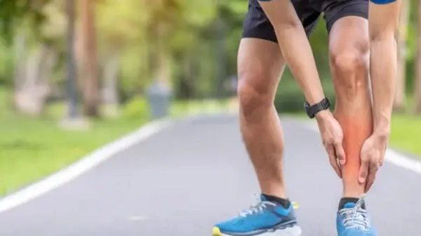
People with shin splints have pain in the bottom third of their shin, which tends to feel like a dull ache like a bruise. It will be become more painful with activities such as running, walking or high impact sports (rugby, soccer, tennis and basketball for example) and will feel better with rest. In the early stages, some people find they are able to push through pain when running only for it to worsen as they continue.


There are a number of other conditions which share symptoms with shin splints; these include stress fractures, chronic exertional compartment syndrome (CECS) or nerve issues, so it’s important to get checked out by a qualified health professional.


Who gets shin splints?

Shin splints is an overload injury – this means that it occurs most commonly when there has been an increase in exercise which is more than the body can handle. This could be someone who has started running and is rapidly increasing their milage, or someone who has been training intensely all season for a sports team. People in the military are also very prone to it due to the high impact that they experience as part of their jobs – up to 35% of military personnel (Moen et al, 2012)!  There is also some evidence that people with flat feet or a narrow running stance are more prone to it (Winters et al, 2018).


What is actually happening?

There are two main theories as to what causes shin splints; one thought is that the bone itself gets overloaded, and this is what causes you pain, whilst the other is that the membrane around the bone is inflamed (Winters et al, 2018).


What does treatment involve?

As shin splints is an overload issue, that means we need to stop the things that are making it worse. Everyone is unique with different activity levels so we’ll work with you to identify your particular aggravating factors. For runners, this might be reducing your weekly mileage and replacing it with cycling to keep your fitness levels up. For people playing rugby, we might focus on drills and skills and less on sprints.


You will also implement exercises to help you get stronger. The stronger a muscle is, the more force goes through the muscle and the less through the bone. Working on the muscles in your lower leg can help with absorbing forces from running and avoiding irritating the sore bone.


It is important to not exercise through pain with shin splints. We know from research that ‘toughing it out’ means it takes a lot longer to heal – runners who ran through pain took, on average, over 100 days to return to sport (Moen et al., 2012). We find people recover better when they progress their exercises without increasing their pain.

Is there anything I can do to prevent it?

Absolutely! One of the most important things is slowly building up activities. For running, we normally recommend only increasing milage by about 10% each week. It’s normal to get occasional twinges but if you notice it continuing or getting worse, come and chat with our friendly team of physiotherapists and we can help you get back on track.

Here are three exercises that can help you prevent shin splints. Aim to complete 3 sets of 20+ a week – when they get easier, add some weight for an additional challenge.


  • Soleus raise – the soleus is one of two calf muscles and is more active when your knee is bent. It is a very important muscle involved in running and is not often trained specifically. This exercise can help target it and make it stronger so that it can keep working for longer.
  • Tibialis anterior raise – your tibialis anterior is a muscle at the front of the shin and helps absorb force as you run.
  • Single leg deadlift against a wall – this works the hip muscles that keep your knees in alignment really well. It also has the benefit of strengthening your hamstrings, which are a common running injury


References

Moen M., Holtslag L., Bakker E., Baten C., Weir A. et al, (2012) ‘The treatment of medial tibial stress syndrome in athletes: a randomize clinical trial’ Sports Medicine, Arthroscopy, Rehabilitation, Therapy & Technology: SMARTT 30 (4)

Winters M., Bakker E., Moen M. et al, 2018 ‘Medial tibial stress syndrome can be diagnosed reliably using history and physical examination’ British Journal of Sports Medicine 52(19) pp.1267-1272

Sciatica Pain

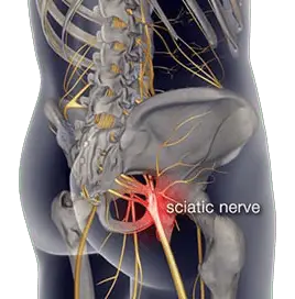
Sciatica is not a diagnosis, it is a term that describes symptoms of pain, pins and needles, numbness and in some cases weakness that radiates along path of the sciatic nerve from the lower back to buttocks and leg.

Causes of Sciatica?

  • Disc derangement / herniation
    Disc injuries are most common cause of sciatica. Discs are cushion like pads located between each spinal segments that act as shock absorbers. The core interior of the discs is made up a gel like substance called the nucleus pulposus surrounded by thick fibrous outer ring called the annulus. Sudden forces applied to the disc can result in the the core interior to push through the outer ring resulting in a disc bulge or in severe cases can rupture the outer ring resulting in disc herniation which can compress the nerve root.
  • Disc degeneration / Arthritis / stenosis
    Age related degenerative changes in the spine can result thinning of the disc and narrowing of the spinal joints. Overtime the narrowing results in bony growths that can compress the nerve roots resulting in sciatica.
  • Soft tissue injury resulting in inflammation
    Thick ligaments and connective tissue envelope the spinal segments to optimize stability. Injury to any of the structures will result in inflammation and swelling which can affect the sciatic nerve directly resulting in sciatic symptoms.
  • Piriformis syndrome
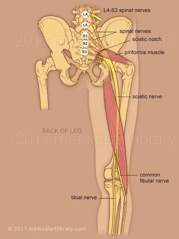
    Piriformis muscle is a located deep in the buttock region. It originates from the sacrum and inserts into the upper part of the hip. The sciatic nerve travels adjacent to the piriformis muscles. Injury to the muscle resulting in tightness or spasm directly affects the sciatic nerve resulting in symptoms.
  • Other possible causes:
    • Sacroiliac Joint Dysfunction
    • Hip joint injury or arthritis
    • Spinal fractures
    • Tumors

Anatomy of the Sciatic nerve

Sciatic nerve is the largest nerve in the human body. It originates in the lower back from five branches of nerves that extend from the spinal cord. The branches exit the spine at nerve roots L4, L5, S1, S2, S3 connect together to form the sciatic nerve.

The large sciatic nerve then travels deep in gluteal region and descends vertically down to the back of the thigh. It supplies motor function and sensation to the skin and all muscles in the posterior compartment of thigh.

At the knee joint the sciatic nerve then divides into two branches the tibial nerve and common fibular nerve.

What exactly does it feel like?

Symptoms of Sciatica are often characterized by one or more of the following features:

  • Unilateral. Sciatica is typically affects one leg.
  • Pain. Nature of pain is often constant with heaviness or dull ache. You may experience sharp, shooting, electric shocks intermittently with postural movements.
  • Neural irritation. pins and needles with occasional postural numbness is common. Postural numbness can occur when you sit or stand for a period of time, but should resolve with movement.  However, if numbness is constant you must be reviewed by your general practitioner or your physiotherapist. 
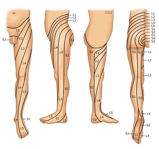
  • Location of pain: symptoms of sciatica are felt along the path of the large sciatic nerve. The following picture shows the potential pathways you may feel your symptoms radiate to depending on the origin of nerve irritation or entrapment. Most commonly the areas affected included the lower back, lateral thigh extending to the calf and foot.

If symptoms are presented on both sides with additional symptoms outlined below – this could warrant an urgent medical review.

Red flags

Signs and symptoms that require prompt medical assessment include:

  • Age >50 years
  • History of trauma
  • Severe unrelenting pain that does not resolve with rest or pain control
  • Partial or complete loss of bowel and bladder function or control
  • Numbness in private regions and the affected side of leg or both
  • Discoloration of skin in comparison to unaffected side
  • Recent or current infection with fever chills and night sweats
  • Sudden unplanned weight-loss
  • History of cancer, kidney dysfunction

Diagnosis

It is important to correctly identify the cause your sciatica is essential in order to formulate an effective treatment plan to manage your symptoms and improve function.

Your GP or a physiotherapist will conduct a thorough diagnostic assessment. Your consultation with your clinician will begin with a comprehensive conversion that allows your clinician to formulate an understanding around potential causes of your symptoms. This includes questions specific to your presenting concerns, general health, history of injuries contributing and medication history and your symptoms management strategies. A physical examination is then followed where by your clinician will assess the range of movement of your lower back and lower extremities, reflexes, strength and sensation assessment to test the integrity of the nerve.

Radiographic examination

Further diagnostic examination in forms of radiographic imaging may be recommended by your clinician to assess the quality of your joints, alignment and check for the presence of any potential lesions contributing to your symptoms.

  • XRAY – commonly used in initial stages to review underlying joint pathology such as wear and tear of joints, fractures or in some cases to view lesions or tumors
  • MRI – high standard imaging that is able to examine in very refined detail possible soft tissues such as muscles, ligaments and internal organs as well as the bony architecture and possible disc injuries.
  • Discogram – A discogram test may be helpful in determining abnormalities in an intervertebral disc. A contrast dye injected into the tissues may allow abnormalities in the disc, such as bulging or herniation to be seen on a medical imaging scan (such as computed tomography scan).

Treatment 

It is advisable to treat sciatica as early as possible in order to avoid the progression of symptoms. Treatments for sciatica may include both non-surgical and surgical approaches.
Typically, non-surgical management is recommended first. Surgery may be required if non-surgical methods have failed to manage your symptoms or the underlying cause is causing deterioration of symptoms. However, in a few severe cases where red flags are presented, surgery may be considered as the first option

Non-surgical approach is the first step to management. This includes intake of pain medications as prescribed by your doctors and referral to physiotherapy.

Pain medications 

Your doctor will prescribe pain medications best suited for your symptoms. These may include

Non-steroidal anti-inflammatory medications such as ibuprofen, celecoxib
Neuropathic medications such as gabapentin, amitriptyline
Analgesics such as codeine, tramadol or oxycodone.
Muscle relaxants such as norflex

Physiotherapy

Physiotherapy will incorporate a combination of gentle strengthening, stretching, and the use of manual therapy to facilitate therapeutic gains.

The goals of physiotherapy for sciatic symptom management includes:

  • Strengthen muscles of the spine, core and lower extremities.
  • Improve flexibility of any tight muscles
  • Improve mobilization of the sciatic nerve
  • Facilitate optimal circulation through slight conditioning exercise (walking, swimming)
  • Education around activity modifications (especially for work-related participation)

Alternative therapies such as acupuncture may be recommended in combination to physiotherapy to facilitate management of your symptoms.

Acute mild sciatica usually improves with 4 to 6 weeks with regular conservative treatment. However, for moderate to severe cases of sciatica especially with a chronic underlying pathology pain may last over 8 weeks and, treatment time may take longer.

Steroid Injections 

Steroid injections are slightly an invasive method used for pain management. Your specialist or an orthopedic surgeon may recommend and administer the injection. In addition to this, injections are also used as a diagnostic method to identify the target nerves or nerve roots affected. The common types of injections for sciatic pain relief include epidural injections.

Surgical approach

In cases where pain and or weakness persists for more than 6-8 weeks or if your symptoms are affecting everyday activities – Surgery may be considered. Your physiotherapist or doctor will arrange the referral for you to meet with an orthopedic back surgeon. Depending on the cause of your sciatica, your surgeon will discuss with you in detail the intended surgical approach, risks involved, post operative management and possible adverse reactions you may have after surgery.