fbpx
Your first step to recovery

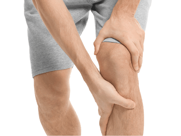
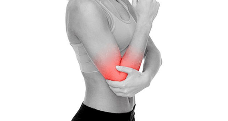
Wrist pain: De Quervains tenosynovitis

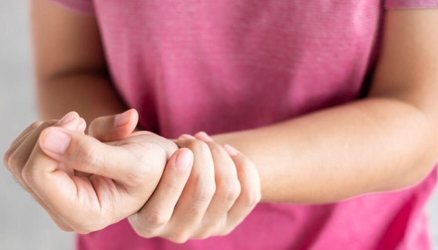
De Quervains tenosynovitis is a painful condition caused by inflammation of two prominent tendons that are located at the wrist and thumb.

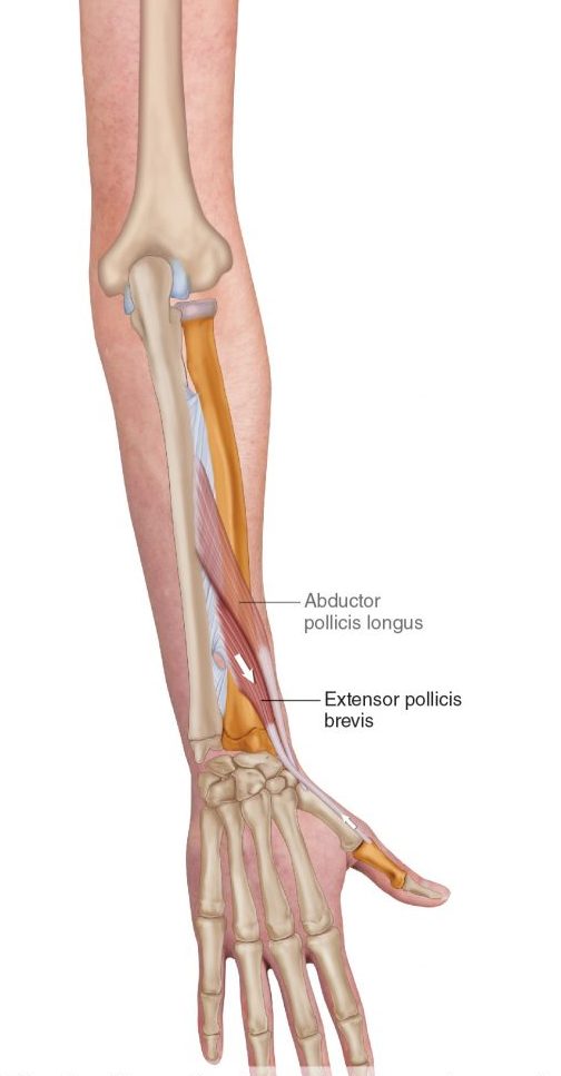
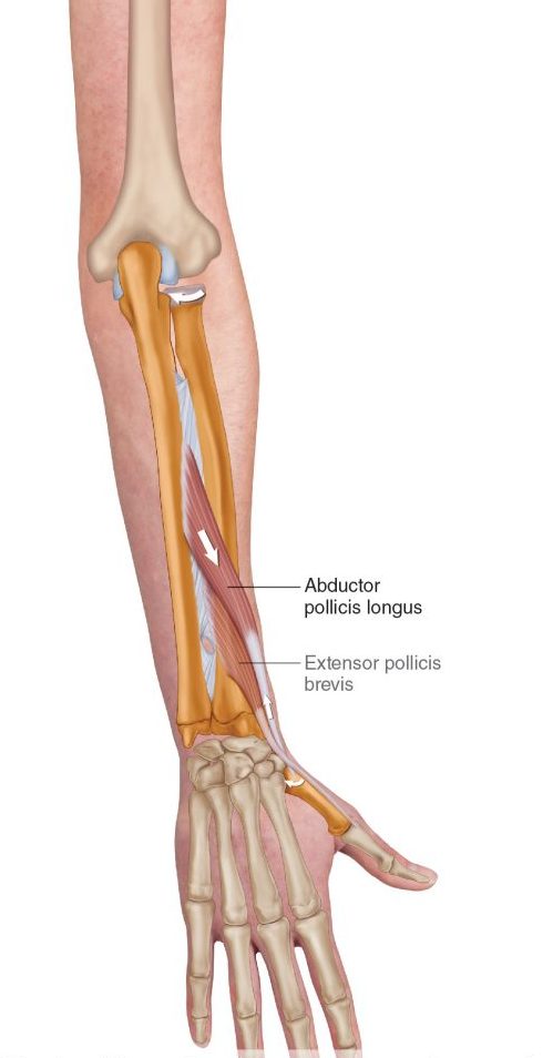
The two tendons called the Extensor pollicus brevis  and Abductor pollicus longus originate from the middle of the forearm travel down towards and over the wrist to insert into the thumb. Collectively they function to extend the thumb, whilst abductor pollicus longus extends and also abducts the thumb (lifting thumb up to the ceiling).

What causes it?

The most common cause of De Quervains tenosynovitis is the repetitive overuse of thumb and wrist whether it is occupational or hobby related. For example, the repetitive thumb movement whilst using scissors by hair dressers, landscapers using shears or whilst gardening). Trauma to the tendons from injuries to the wrist or the thumb can cause inflammation of the tendons.

In some cases, age related degeneration of the tendon sheath or underlying conditions such as rheumatoid arthritis increases the risk of the developing De Quervains tenosynovitis. Hormonal changes resulting in fluid build up in young mothers can commonly result in De Quervains tenosynovitis.

Symptoms

Commonly your symptoms may include:

  • Pain located at base of your thumb
  • Pain elicited by movement of thumb (gripping or making a fist)
  • Grating or snapping feeling
  • Tightness in the wrist
  • Swelling surrounding the base of thumb and wrist

How is De Quervains tenosynovitis diagnosed?

Your doctor or physiotherapist will be able to diagnose the condition based on your symptoms and after doing a thorough movement assessment to rule out any other potential diagnosis.

  • Finkelstein test is used to elicit symptoms to confirm De Quervains tenosynovitis.

How to test:

  1. Wrap your thumb with your fingers.
  2. Slowly bend your wrist down

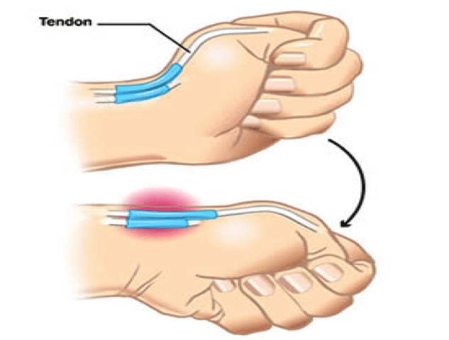
A positive test would elicit pain at the site of the two tendons.

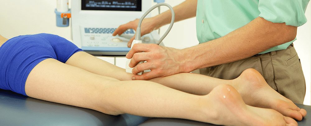
Radiological investigations in lights of ultrasound and an x-ray might be recommended for further investigations, particularly to confirm clinical diagnosis or to rule out any other possible causes of De Quervains such as osteoarthritis.

What treatment options are available?

Conservative (non-surgical) management

Conservative management measures are generally recommended as the first line of management for mild to moderate symptoms. This is because up to 60-70% of symptoms are likely to improve over a period of 6-8 weeks of regular physiotherapy intervention. In this period, the following strategies are recommended by your therapist to fast-track your recovery

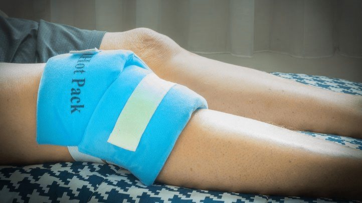
  • Rest and application of heat or cold packs
  • Avoid repetitive use of thumb
  • Pain medications (anti-inflammatory medications) such as diclofenac or ibuprofen
  • Splints or braces
  • Steroid injection

Surgical management

In more severe cases when conservative management has failed, surgery may be recommended by an orthopaedic specialist or surgeon.

Prior to your surgery you will have the opportunity to thoroughly discuss with your surgeon the details of the surgical procedure and about the post operative rehabilitation process.

  • Surgical procedure

Surgery may be performed under general or local anaesthesia. A small incision is made at the wrist and thumb region. The covering of the tendons (sheath) is then separated and expanded to provide the tendon space to allow the tendon to move smoothly within the sheath. After this the, the incision in then sutured with a firm dressing applied over the suture site.

  • While you recover from the surgery, an information sheet with post operative guidelines will be provided to you by your surgical team. It is important that you must follow the guidelines recommended by your surgeon for optimal recovery.
  • In most cases your will have a follow up with your surgeon few weeks after your surgery to check your wound healing and your progress. You are often times referred to physiotherapy for strength and conditioning of your wrist and hand movements to facilitate your recovery.

Repetitive Strain Injury (RSI) Explained


RSI is typically defined as an overuse disorder- a gradual build-up of overload to nerves, tendons, and muscles arising from repetitive movements or activities. Repetitive use of the same motions leads to inflammation and damage to these soft tissues. This disorder mostly affects the upper limb- particularly the elbows, hands and wrists.


Causes

Possible causes of RSI include but are not limited to:

  • Undertaking the same and repetitive movements and stressing the same muscle groups
  • Working in cold environments
  • Assuming a sustained and/or awkward posture for prolonged periods of time
  • Undertaking a particular activity for prolonged periods of time with no rest-breaks
  • Frequent and prolonged use of vibrating equipment
  • Adopting poor postures from working at inappropriately designed workstations
  • Undertaking a motion which involves carrying and/or lifting heavy items


Symptoms

RSI leads to a gradual development of a broad variety of symptoms, which range from mild to severe in severity. RSI particularly affects the muscles and joints of your wrists, hands, elbows, forearms, shoulders, neck. Having said this, RSI can affect other areas of the body as well.

Common symptoms may include:

  • Pain
  • Tingling
  • Cramping
  • Increased sensitivity to heat and cold
  • Tenderness
  • Fatigue
  • Loss of strength
  • Throbbing
  • Soreness
  • Achiness
  • Stiffness
  • Struggling with typical activities of daily living, such as gripping and twisting motions, carrying light weights, writing, kitchen prepping, dressing, personal cares etc

You may develop these symptoms when you undertake a task repetitively for a period of time, and can settle when you stop. Symptoms may settle over a few hours or over the course of a few days. However, if left untreated or is poorly managed, a minor RSI may gradually progress to a nasty chronic injury.


Diagnosis


If you experience mild discomfort whilst completing particular activities at home or at your job, it is a good idea to see your GP or physiotherapist to talk about RSI. But an RSI is not always simple to diagnose as there is no particular clinical test for it. Your GP will enquire about your medical history, occupation and work environment, and other activities to attempt to identify any repetitive motions you undertake that may be the cause of your symptoms. A physical examination will be undertaken, where they will assess your movement, check for pain, inflammation, sensation, tenderness, strength and reflexes in the impacted body part. RSI may be triggered by specific health disorders like bursitis, carpal tunnel, tigger finger, ganglion cyst, or tendonitis (inflammation in your tendons). Your GP can refer you on further diagnostic tests such as X-rays, Ultrasounds, blood tests, MRIs, nerve conduction tests etc, to determine if these underlying disorders may be the cause of your symptoms. You may be also be referred onto a physiotherapist and acupuncturist for conservative treatment and management for mild-moderate issues. If symptoms persist, you will then be referred onto a specialist.


Management

Initial treatment options for the management of RSI symptoms is conservative. This includes:

  • Rest, Ice, Compression, and Elevation (RICE principles)
  • Taking regular breaks between tasks and looking after your posture
  • Undertaking your activities and movements with appropriate form and posture
  • Intake of Nonsteroidal anti-inflammatory drugs (NSAIDs), both oral and topical as prescribed by the GP
  • Use of cold and heat to the impacted area
  • Administration of steroid injections into inflamed joints and tendons
  • Tailored exercise prescription from physiotherapists to correct posture and strengthen and stretch affected muscles
  • Acupuncture
  • Stress reduction and relaxation training
  • Use of splints and braces to help protect and rest the affected muscles and tendons

Ergonomically appropriate adjustments to your workstation and work environment may be recommended by your physio and GP- for example resetting your desk and chair if you’re working at computer, and alterations to your equipment and activities/motions to lessen the strain and stress on your muscles and joints. Surgery may be necessary in some cases.


Prevention

Minimizing repetitive actions particularly if they involve the use of heavy machinery or vibration. Improving your working posture and work-environment as well a taking regular breaks. Employers often undertake risk-assessments when you join a company to determine that the work area is ergonomically fit, comfortable and appropriate for you. You may be able to request for an assessment if you have not had one or are having issues with your work environment

Tennis Elbow Explained

What is Tennis Elbow?

Tennis elbow, or also known as lateral epicondylitis, is a painful disorder of the elbow triggered by overuse. This disorder is characterised by inflammation or, in some cases, micro-tearing of the extensor tendons which link the forearm muscles on the outer part of the elbow. This leads to pain and tenderness on the outside of the elbow, spreading through to the forearm and wrist.

Elbow Pain - Do I have Tennis Elbow? - Oh My Arthritis

Causes

Tennis elbow is characterized as an overuse and muscle strain injury. It is caused by repetitive and/or vigorous contractions of the forearm muscles which is used to straighten, extend, and lift the wrist and hand. The repetitive motions and stress to the tissues can result in a string of tiny, microscopic tears in the tendons which attach your forearm muscles to the bony prominence on the outside of the elbow.

Tennis elbow may result from number of activities including but not limited to:

  • Typing and repetitive computer mouse use
  • Cutting/chopping motions
  • Manual work that involves repetitive turning or lifting of the wrist, such as plumbing, or bricklaying.
  • Gardening
  • Using hand tools such as scissors, clippers, screwdrivers, plumbing and carpentry tools
  • Playing racquet sports, such as tennis, badminton, or squash
  • Throwing sports, such as the javelin or discus
  • Painting
  • Sewing/knitting

Tennis elbow may also occur after a sudden knock or bang to the elbow, if you undertake activities that you are not used to excessively and aggressively, and sometimes there is no apparent cause for it.

Risk Factors

Factors which may increase your risk of tennis elbow include:

Your age: Whilst this disorder impacts people of all ages, it is mostly prevalent in adults in the age group of 30-50 years old.

Your occupation: Those who have jobs which entail repetitive movements of the wrist and arm are more likely to develop this disorder. Examples include but are not limited to painters, computer users, plumbers, butchers, carpenters, and chefs.

Sports: Partaking in racket sports increases the risk of tennis elbow, particularly if you use poor form and technique.

Common Signs and Symptoms

Tennis Elbow - Symptoms, Causes, Treatment & Exercises

Pain noted around the bony knob on the outside of your elbow is the most common characterising symptom of tennis elbow. This knob is where the injured tendons connect to the bone. The pain is often depicted as “burning” in nature. Your elbow may be tender and sore to touch, and the pain can refer down to the forearm. The pain often increases with gripping, grasping, or rotating motions of the wrist and forearm. Bending and straightening your elbow may also be painful.

The severity of your pain may vary from a mild discomfort to severe pain that can interfere with your sleep and day to day activities. The pain typically starts gradually and then worsens over weeks or months.

Diagnosis

During your physical examination your physiotherapist will attempt to produce your pain in your elbow via specific tests and movements. They will assess your range of motion in your elbow, wrist, and shoulder joints. Referrals for X-rays and ultrasound scanning may be indicated to further support your diagnosis and to rule out other potential sources of your pain

Management

A mix of non-surgical treatment options are effective for the majority of tennis elbow cases, and self-resolves over time. You should rest your elbow and painful activities should be avoided. But it is very vital to maintain gentle movements of the forearm, elbow, and wrist through its range of motion.

Potential treatment options include:

  • Ice
  • Rest
  • Physiotherapy and acupuncture
  • Anti-inflammatory medications as recommended by your doctor or pharmacist
  • The use of a wrist and forearm brace or splint to support and rest your forearm

As your initial elbow pain lessens, your muscles around the elbow, forearm and wrist should be safely strengthened and stretched under guidance of a physiotherapist. Your physiotherapist will advise you on particular exercises, give you appropriate symptom management advice and take you through a personalised graduated rehabilitation program. If you continue to experience pain after 6-8 weeks of treatment, your physiotherapist can refer you back to your doctors, to consider administration of a cortisone injection into the elbow to help reduce pain and inflammation, and further referral onto see a specialist to seek guidance on other treatment options.

Helpful Exercises

KNEE PAIN

Knee pain is one of the most common musculoskeletal complaints that affects peoples of all ages.
Knee pain can result from injuries of traumatic nature or due to complications from medical conditions.

Depending on the structures involved, pain can be localized to a specific area or be felt all
around the knee.


ANATOMY OF KNEE

The knee joint is a hinge joint. Other than bearing the weight of the body, it’s primary function is to bend, straighten and rotate to a small degree. To achieve this function, the knee joint relies on a number of structures.


Image result for knee anatomyImage result for knee anatomy


Bones

Knee joint consists of four bones to provide structure and weight-bearing ability.

  •  Lower end of thigh bone (femur)
  •  Upper part of shin bone (tibia)
  • Knee cap (patella)
  • Fibula (not involved in weight-bearing, but provides attachments for ligaments and tendons)


Ligaments

Four important ligaments connect the two big bones, providing multi-directional stability.

  • Cruciate ligaments

Anterior cruciate ligament (ACL)

Posterior cruciate ligament (PCL)

  • Collateral ligaments

Medial collateral ligament (MCL)

Lateral collateral ligament (LCL)


Cartilage

  • Glossy cartilage lines the end of each bone to protect and allow smooth movements against each other with almost no friction.
  • Meniscus is another type of strong cartilage that lines the upper surface of the tibia bone to cushion and stabilize the knee.


Tendons

There are two important tendons located on the front of the knee joint.

  • Quadriceps tendon is a strong durable tissue that extends from the quadriceps muscle and connects it to the knee cap.
  • Patella tendon connects the knee cap to the tibia bone.


Bursa

Bursa are fluid filled sacs that are found in areas that require the most protection. They occur where ligaments, muscles, skins, tendons or bones rub together.


Muscles

Many muscles cross the knee joint, some of which cross from the hip or ankle joints. Due to this, some people may experience knee pain as a result of muscle imbalances such as weakness, poor flexibility and or dynamic control.


CAUSE OF PAIN

Some of the common causes of knee pain include:

  • Ligamentous sprain
  • Meniscus injuries (bulge or tears)
  • Fracture or dislocation
  • Tendinitis, Sprains
  • Bursitis
  • Calcification
  • Baker’s cyst
  • Arthritis (osteoarthritis)
  • Overuse syndromes (patella-femoral syndrome, chondromalacia)
  • Autoimmune conditions (Infection, Gout, Lupus, rheumatoid arthritis)


WHEN TO SEEK HELP

Consult your doctor or physiotherapist if your symptoms have not subsided after one-week of consistent self-management (RICE, pain medications or alternative pain-relieving modalities), or if your knee pain is stopping you from managing your hobbies or day to day activities.


Immediate medical attention: 

Knee pain from with the following signs and symptoms may require immediate attention:

  • Severe pain
  • Pain that does not resolve with rest
  • Sudden swelling or bruising
  • Clicking or locking of the knee
  • Inability to bend or straighten the knee
  • Inability to weight bear


SIGNS AND SYMPTOMS

  • Pain
  • Swelling
  • Bruising
  • Stiffness
  • Clicking, locking
  • Redness


DIAGNOSIS:

Treatment of your knee pain will depend on its underlying cause. So, it is all about the diagnosis.

A focused subjective and physical examination of your knee will be performed by your physiotherapist.


Subjective

Your physiotherapist will ask a range of questions

  • Location of pain – front or behind the knee
  • Description of pain – dull ache or sharp
  • The behavior of pain – constant or intermittent
  • Aggravating and easing factors
  • General health
  • Goals of treatment


Physical assessment

Your physiotherapist will inspect your knee joint to diagnose the source and the potential underlying cause(s) of pain.

You may be referred to have radiological Imaging to make or confirm the diagnosis.


Radiology

Image result for xray knee


TREATMENT

In most cases, individuals suffering from knee pain respond well to conservative modes of treatment (pain relief, physiotherapy, acupuncture, etc). Surgical intervention may be required where conservative management has failed to optimize function and reduce pain.


CONSERVATIVE MANAGEMENT

A self-management remedy to control inflammation (pain and swelling) in acute or chronic knee pain is using the ‘RICE’ principle (rest, ice, compress, elevate)

  • Rest – refrain from activities that impose repetitive strain or aggravation of knee pain
  • Ice – use an ice pack for 10-15 minutes, 2 to 3 times per day (with care)
  • Compress – use a compression bandage to reduce swelling (not to be worn when sleeping)
  • Elevate – using pillows elevate injured leg. This works best when the leg is higher than the level of heart, to use gravity to help facilitate the circulation of fluid.


Pain medications

Over the counter pain relievers such as non-steroidal anti-inflammatory medications (ibuprofen, celecoxib) play an important role in reducing inflammation and pain.

(Note: If you have problems with bleeding, stomach ulcers or other liver, kidney conditions, anti-inflammatory medications MUST NOT be consumed without consulting your doctor. 

Visit your general practitioner for more information on what medications are right for you.


Physiotherapy

After establishing your diagnostic findings, your physiotherapist will devise a tailored recovery programme to help you manage your pain, improve strength and flexibility.

Your physiotherapist will work with you to advance your understanding of your symptoms and provide a range of exercises, stretches and self-managing strategies that will help you be in control of your recovery.

As required, your therapist may liaise with your doctor or other health professionals (acupuncturist, podiatrist, knee specialists) to facilitate your progress.


Acupuncture/Acupressure

Acupuncture and acupressure are two different options available for individuals suffering from pain and swelling. While both aim to help control inflammation and fasten healing and recovery, acupuncture involves inserting thin needles into the body, whereas acupressure relies on hand pressure and some forms of massage.


Steroid Injection

In some instances, knee injections are recommended by your physiotherapist or doctor to reduce inflammation and relieve pain.


SURGERY

Surgical intervention may be required where conservative management has failed to optimize function and reduce pain. In this case your physiotherapist will refer you to a surgeon for the opinion of care.


EXERCISES FOR KNEE PAIN

The thought of exercise when you have knee pain can be daunting. However, your trusted physiotherapist will work with you to provide specific ‘pain-free’ exercises to get you started on effective strengthening.

Image result for knee exercise cycling

Alternatively, low-impact activities such as cycling or elliptical machines are great. Notice what feels right for you. Swimming, jogging in water, or water aerobics may be appropriate if skin integrity is maintained.

Note: muscle soreness after a hard workout is normal. 

If you experience sharp, shooting, or sudden knee pain you must consult your physiotherapist or doctor.

5 COMMON MISCONCEPTIONS ABOUT PHYSIOTHERAPISTS – PART 2

Myth 1: When you see a physiotherapist you just lie on the bed and get given an ice or heat pack.

At Physio Fusion we use an active approach to treatments. Physiotherapy will include manual hands-on therapy to facilitate tissue healing and tissue load tolerance alongside an exercise program individualized to your needs.

Myth 2: If I have elbow pain then the injury must be in my elbow.

The area of pain is not always the area that is the issue! It may be a result of a previous injury that was never fully rehabilitated. This is where we can help you out. Our assessment will consist of gathering information on your presenting complaint, any previous injuries or traumas (physical and emotional) and a medical history followed by an objective evaluation of your body. This allows us to get to the root cause of the pain and manage your symptoms most effectively.

Myth 3: I can’t do any of my normal activities while I attend physiotherapy.

Not true! Our Physiotherapists want to keep you as functional as possible whilst allowing your injury to heal. During the initial assessment your physiotherapist will determine what activities you can do and advise you on those that must be avoided. You will then be given clear and timely objectives to ensure you reach your goals to get you back doing what you love!

Myth 4: A scan will show me exactly what is wrong.

Sometimes it will, but sometimes it won’t. It’s no secret that our bodies change as we age, so even people without pain are likely to have an imperfect scan. Medical imaging can sometimes play an important role in the assessment and management of your musculoskeletal issue. When necessary, your physiotherapist will know what type of imaging to refer you for.

Myth 5: Is cracking my back/neck/knuckles bad for me?

There is no strong evidence to suggest that ‘cracking’ your joints causes degeneration, laxity or instability. The ‘cracking’ occurs when we move a joint to its end range. The audible sound happens because of ‘cavitation’ in the joint; this involves gas bubbles popping within the fluid surrounding the joint as pressures change.

But is it good to crack?

Self manipulation can be a helpful way to reduce the feeling of stiffness or tightness. If you are finding that you need to ‘crack your joints’ often it is good to know that there are many other more beneficial ways to provide greater long term relief.

Top tips:

  1. Ask your physiotherapist to provide you with some specific exercises to help you overcome the feeling of stiffness or tightness.
  2. Move regularly and avoid movements or positions that exacerbate your symptoms until you have been seen by your physiotherapist.

5 COMMON MISCONCEPTIONS ABOUT PHYSIOTHERAPISTS- PART 1

Myth 1: ‘’Physiotherapy is just massage’’

This is a common phrase people use to describe a physiotherapists role. It is true that as a profession we like to work ‘hands on’. This is because our profession is directly affiliated with the anatomy of our clients and by using touch we can more accurately assess, locate and treat any dysfunctional motions within your body. Our physiotherapists like to use massage as it is a great way reduce pain and improve bodily function but their skill set is much more extensive than this.

Did you know?

There are well over 20 different treatment approaches commonly used by physiotherapists.  These may include, but are not limited to:

  • Exercise prescription.
  • Joint mobilization.
  • Joint manipulation.
  • Instrument mobilization.
  • Muscle energy techniques (improves muscle and joint function).
  • Neurodynamics (mobilization of the nervous system).
  • Taping.
  • Dry needling.
  • Acupuncture.

Myth 2: Always sit up straight! Slouching is bad.

In fact, forcing yourself to adhere to the traditional ‘good posture’ (back straight and shoulders back) may be putting unnecessary tension on your body. That isn’t to say being upright is bad either, but in life we must always find a balance. To reduce stress on your body whilst seated, ensure your back is supported and your feet firmly on the ground. To prevent and reduce the likelihood of back pain we must look to vary our posture frequently throughout the day and ensure we take short breaks to stretch, stand and walk.


Myth 3: Physiotherapy sessions are painful

Physiotherapists aim to help you reduce pain and get you back to doing what you love! Whether your injury is acute or chronic our physios always ensure they work within your pain threshold to help you regain lost movement and function.

Myth 4: I need a referral to see a Physiotherapist’

We have got you covered!

Remember, physios are registered health care professionals. Physio Fusion is a registered ACC provider. This means that if you have had a recent injury (within one year), you can see us directly and we can help you lodge an injury claim directly with ACC. You do not need a referral for your doctor.


Myth 5: Lower back pain …. surgery is my only option

Around 70-90% of the total world population will experience lower back pain during the course of a life time. Symptoms can range from mild to severe and can either last for short or long periods or remain constant. Back pain can be very debilitating and when the pain does not resolve as soon as anticipated many fear that surgery may be the only option. In few cases surgery may be necessary, but for the majority there’s often nothing to fear, and with time and a gradual loading or exercise program, you can make a full recovery.

Did you know ?

Back pain is not always a sign of injury or damage. Each individuals back pain story will be different, and for many non-physical factors play a huge part in their story. These factors can be:

Psychological –

  • Stress.
  • Fear of movement.
  • Depression.

Health related –

  • Sleep deprivation.
  • Physical inactivity.
  • Smoking (nicotine decreases blood flow to your back).
  • Overweight.

Physio Fusions top recommendations for keeping back pain away:

  • Keep active – regular exercise nourishes joints, strengthens muscles, increases blood flow and improves your mood.
  • Sleep well.
  • Maintain a healthy weight by ensuring you maintain proper nutrition and diet.
  • Sit in a chair with good lower back support.
  • Quit smoking.
  • Ensure that when you lift or pick up objects you do so in a safe manner. This is something one of our physiotherapists can guide you with.

See a doctor urgently if you have the following symptoms:

  • Numbness in the groin or buttocks.
  • Loss of bladder or bowel control.
  • Redness or swelling on your back.
  • Difficulty walking.
  • Constant pain, especially at night.
  • Pain that is getting much worse, or spreading up your spine.
  • Numbness or pins and needles in both legs.
  • Feeling unwell with your back pain, such as a fever or significant sweating that wakes you from sleep.

INVEST IN YOUR HEALTH NOW OR PAY FOR YOUR HEALTH LATER!

It is scientifically proven that physical exercise is one of the greatest rehabilitation methods. Numerous studies consistently prove the indisputable benefits of exercise therapy.

Did you know? 

People who are physically active for approximately seven hours a week are 40% less likely of dying early than people who are only active for less than 30 minutes a week.

So what are the health benefits of exercise?

  • Helps you control your weight.
  • Can help you develop positive and healthy social relationships  (making friends, engaging with other children, self-expression).
  • Improves coordination and balance.
  • Improves posture.
  • Strengthens your bones and muscles, slowing down degenerative processes such as osteoarthritis.
  • Reduces your risk of falls.
  • Reduces your risk of some cancers, including breast, lung and colon.
  • Improves your mental health and mood.
  • Improves your sleep.

Any many many more!!

So what is stopping you?

Time – Time waits for no one. But there is plenty of it in the week to make a difference. Don’t fear moving slowly forward, fear standing still.

Knowledge – Starting is always the hardest part! Here at Physio Fusion we are always willing to help guide and advise you on all aspects of your physical health. We are very fortunate to have good connections with other healthcare providers who can also help to assist you!

Confidence – You have our vote! At physio fusion we embrace the lifestyle we promote. We are here to facilitate your needs and to guide and advise you on all aspects of your health.

Cost – Your health is your wealth. At Physio Fusion we provide the highest quality treatment and advice at very affordable prices.We have special rates for clients with a Gold Card (no co-payment) or Community Services Card ($10.00).

Distance – For those of you unable to travel to our clinics, or who are isolating currently due to COVID-19 we have you covered! We offer telehealth (online video) consultations, supported with a rehab exercise programme tailored to your needs via the online database Physitrack.

Secure your appointment today by booking online or phone us on:

09 626 6186 (New Windsor)
020 417 32135 (Henderson)